Parts of the brain
The forebrain
The forebrain is composed of the telencephalon and the diencephalon. It handles everything which makes us “us” – or at least gives us that impression – including perceptions, consciousness, cognition and voluntary action. The figure below recalls the parts of the brain in development.
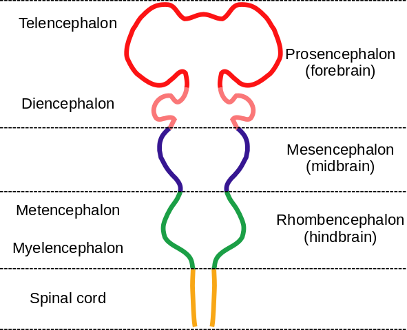
Embryonic brain parts, by “Nrest” via Wikimedia Commons
The telencephalon
The telencephalon develops into the cerebrum, the largest part of the human brain, which consists of the cerebral cortex and several elements below it such as the hippocampus, the basal ganglia (or, more correctly but less historical, basal nuclei) and the olfactory bulb.
The cerebral cortex
The cerebral cortex is essential for all sorts of processing of sense data and motor control. It is where the reasoning and cognition specific to humans (and, to a lesser degree, some other animals) takes place, and is the seat of planning and language, volitional behavior and conscious perceptions, thinking and memory. It is the command center where input sensory information is translated into output motor control. In evolutionary terms, it is the most recently developed part of our brains and has taken over or added to function of older structures.
Its unique and fairly uniform structure (sometimes termed “canonical”) allows for great plasticity in functioning. It consists of layers[ref]Six, in the striate cortex.[/ref] of gray matter (neuron cells, dendrites and synapses) on the outside (distally) and white matter (axons) beneath, although the difference in colors is less pronounced than those terms may imply. It consists of two lateral hemispheres joined by a bundle of axonal connections, the corpus callosum.
Usually, the left hemisphere handles sensory input from and motor output to the right side of the body and vice versa. The left hemisphere handles more of language and detailed analysis than the right. The right side handles visual pattern recognition and overall perception, and seems to be important for the appreciation of music. But the brain is quite plastic (capable of changing), so any strict distinctions between the function of left brains and right brains must be considered with some skepticism.
 |
 |
|
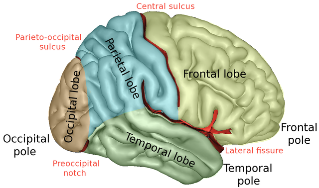
The brain lobes, by Sebastian923 from Wikimedia Commons |
Some important brain structures, from Openstax Biology |
The cortex consists of five lobes partially separated by the sulci and notches. Functions are distributed among the different lobes as follows.
- The frontal lobe, which is located anterior to the central sulcus and above the lateral fissure, is the site of planning and takes into account goals and environment. It is important for movement, as it includes the primary motor cortex. It is also part of the neural circuitry for language and working memory. The activity of this lobe goes from more abstract to more specific (motor) from front to back. The frontal lobe includes a motor map (explained later).
- The parietal lobe, posterior to the central sulcus and above the lateral fissures, receives skin data – touch and pressure – which are mapped here. It also assembles information from other senses, like space around the body, and decides where to direct attention.
- The temporal lobes, on the sides below the lateral sulci, handle auditory processing and visual interpretation, do pattern recognition and are essential to language comprehension and speech.
- The occipital lobe, at the back, contains the visual areas (V1, V2, …) which analyze aspects of sight, such as color, depth and motion. There is a retinotopic map on V1 (explained later).
Some parts assure more specific functions:
- The lateral prefrontal cortex is essential to rationality and planning. It contains short-term memory such as working memory. This is transferred to long-term memory through the hippocampus during REM (rapid eye movement) sleep.
- The cingulate cortex is an evolutionarily older cortex below the neocortex and is part of the mesocortex. It connects the limbic system and the neocortex. Its forward (“ventral”) part, the anterior cingulate cortex, which contains the anterior cingulate gyrus, is important in control by the neocortex, telling it when things are not going according to plan.
- The primary motor cortex controls voluntary movements by means of axons which run down the spinal column and connect to motor neurons at the neuromuscular junction, as we have seen in a preceding chapter.
Sub-cortical structures
These structures lie below the cortex.
- The hippocampus receives input from sensory areas and from the rest of the neocortex and, along with other structures deep within the temporal lobes, such as the rhinal cortex, is essential for conversion of short-term to long-term memory. It has connections to and from almost all the neocortex. By projecting the connections it receives back to the neocortex, it activates the same regions which were originally activated by the senses and so forms a mental representation of the sensory perception.
- The amygdala has a function similar to that of the hippocampus but concerns events of high emotional content, such as moments of fear. Such memory provides for fast, low-resolution, autonomic responses to recognized stimuli, such as odors or the need for flight from dangerous situations. The amygdala works with the orbitofrontal cortex (OFC) to assess risks and rewards and to choose appropriate behavior and remember it. These memories are our emotions and may give rise to what we call intuition. Although the amygdala and hippocampus perform similar functions, they have different inputs and outputs.
- The basal nuclei (formerly called basal ganglia) are hidden deep under the cortex, basal meaning deep within the brain. They are important in planning and executing motor movements initiated by the neocortex. They consist of the caudate, putamen, globus palladus, substantia nigra and subthalamic nucleus. The caudate and putamen together constitute the striatum.
The diencephalon
The diencephalon lies beneath the neocortex and consists of the thalamus and hypothalamus and parts of the so-called limbic system.
- The thalamus is an extremely important double structure which is the gateway for sense data (except olfactory) as well as signals from the rest of the nervous system; it relays the information to appropriate areas of the neocortex.
- The hypothalamus controls the autonomic nervous system. It assures homeostasis by producing hormones which regulate essential functions (temperature, blood pressure, hunger, thirst, circadian rhythm, etc.). As has been discussed already, it is the brain’s gateway to the endocrine system via the pituitary gland, the posterior pituitary being an extension of the hypothalamus. The hormones it secretes include oxytocin, which is released by the posterior pituitary gland and plays a role in caring and childbirth, and vasopressin, which regulates water retention and may also play a role in social behavior and bonding.
The limbic system is an evolutionarily old set of structures which existed before mammals. The term limbic system is historical and does not refer to an integrated structure, so it is often denigrated. It is subcortical (lies beneath the cortex) and is considered to be constituted by a number of structures, including the thalamus, hypothalamus, cingulate gyrus, basal ganglia and the following:[ref]The term “limbic system” is historical and its members are not all in the same embryonic structure. I am not quite certain where they should be housed according to modern standards.[/ref]
The midbrain
Nuclei (groups of cell bodies) in the mid- and hindbrain play a role in essential, non-cognitive functions such as respiration, blood circulation and level of consciousness. They are phylogenetically very old and exist in all vertebrates.
The midbrain, or mesencephalon, includes the following structures.
- The substantia nigra (also considered one of the basal ganglia) is involved in the initiation and maintenance of voluntary movements. Damage to it is linked with Parkinson’s disease.
- The inferior colliculus receives audial information and relays it to the thalamus.
- The superior colliculus receives visual information from the eye and, in turn, controls eye movements (saccades). It holds maps of the visual world. The superior and inferior colliculi together form the tectum, the top of the midbrain.
- The reticular formation runs from the midbrain into the spinal cord. It has a number of different modulatory functions, including control and regulation of the brain’s state of arousal and consciousness.
- The diffuse modulatory system consists of the locus coeruleus, raphe nuclei, substantia nigra and ventral tegmental areas. These cells perform regulatory functions as each of them sends small amounts of a specific neurotransmitter across wide areas of the brain or spinal cord. They can be thought of as a kind of volume control.
The hindbrain
The hindbrain, or rhombencephalon, supports vital processes.
- The medulla is a communication path for axons between the brain and spinal cord and is the place where axons to or from the rest of the body change sides (decussation). It controls automatic functions such as breathing, heartbeat, hearing, touch, taste and swallowing.
- The pons is anterior to the medulla and handles visceral functions and facial expressions. It contains centers like the vestibular nuclei, which receive information about orientation of the body relative to gravity and acceleration, so it is partly responsible for balance. As its name implies, it forms a bridge between the cerebral cortex and the cerebellum.
- The cerebellum (“little brain”) is the most complicated part of the hindbrain. It consists of two lobes at the back of the brainstem. In terms of size, it is second after the cerebral hemispheres. It receives input about the body from the spinal column and about goals of movements from the cortex and coordinates the two. It also memorizes information needed for complex motor skills. Unlike the cerebral cortex, each side controls its own side of the body. Without the cerebellum to modulate and coordinate motor behavior, you would not be able to ride a bicycle – or walk. The cerebellum is the oldest part of the brain from an evolutionary point of view and is necessary to all animals, at least to vertebrates.
The spinal cord
The spinal cord manages basic life-critical functions such as reflexes, breathing or heart rate; its actions are usually unconscious.
As already explained, the spinal cord is segmented, each segment having two pairs of ‘roots’ connecting the spinal cord to other parts of the body.
- The ventral roots are outgoing (efferent) motor-neuron axons, clustered in functionally related groups. Their cell bodies are within the spinal cord.
- The dorsal roots are the incoming (afferent) sensory-neuron axons. Their cell bodies are in dorsal root ganglia, near the spinal cord.
Interneurons may connect the incoming and outgoing circuits so as to allow rapid reflex reactions, like the knee jerk. They also relay commands coming from the brain.
Now go on to study brain inputs — the sensory systems, maps and vision.