Learning and memory

Memory is stored in the brain by means of neural connections (circuits) and so is based on synaptic weights – relative strengths of synapses. These are not programmed in our genes, as there are not nearly enough genes to contain such an immense recipe. Instead, synaptic enhancement is due to correlated activity brought about by chemical substances between the pre-synaptic and post-synaptic elements. This method has the advantage of allowing environmental influence on learning.

It hardly needs to be pointed out that memory is essential not just to what we are, but to our very survival. The “present” we inhabit is really a collection of memories, some from the last milliseconds (what we see as “present’), filled out by and compared to memories of similar past experiences. It really is the “remembered present”.1Edelman (2006).

Types of learning and memory

Neuroscientists and psychologists distinguish several types of memory, which have different functions and are handled and stored differently in the brain.

  • Explicit or declarative memory is conscious. It starts out as short-term memory in the pre-frontal cortex and may later be stored as long-term memory via the hippocampus. Explicit memory in turn is divided into two parts.
    • Semantic memory refers to remembered facts.
    • Episodic memory refers to remembered experiences, events and episodes.
  • Implicit or procedural memory is unconscious. It contains what is necessary for all those procedures we do without thinking, like riding a bike or driving a car, playing a musical instrument or tennis. It is in fact a collection of different systems and depends on the amygdala (emotional responses), the cerebellum (motor control) or the striatum, a part of the “reward system” and input to the basal ganglia (procedural memory).

In addition, memory can be short-term, one type of which is working memory, or long-term.

Scientist also study several basic types of implicit or procedural learning.

  • Non-associative learning describes memory due to a single stimulus. The two types of non-associative memory are:
    • Habituation is just that, getting used to a stimulus (such as background noise) so you stop paying attention to it.
    • Sensitization is the opposite of habituation, like “getting burned” once so any heat frightens you unduly afterwards.
  • Associative learning forms associations between two events and also exists in two types.
    • Classical conditioning is the association of a neutral stimulus (like a bell ringing) with a real or effective one (the smell of food). The effective response is called the unconditional stimulus (US) because it works all the time; the learned one is called the conditional stimulus (CS). Such conditioning is effective only if the CS occurs at the same time as or slightly, but not too much, before the US.
    • Instrumental conditioning is associating a stimulus with a reward, a behavior with a result.

Now let’s look at mechanisms of learning and memory.

Simple implicit learning in invertebrates

Several mechanisms for learning have been identified, all based on the basic fact of learning and memory, which is this:

Neural circuits, the basis of what goes on in the brain such as learning or plasticity, are set up by modification of synaptic strengths.2I see it as the enhanced synapses’ having less resistance to passage of electrochemical neuronal circuits, so that signals take the path of least resistance from one neuron to another. Physicists like minimizing paths.

This synaptic modification depends on correlated activity in pre- and post-synaptic terminals. The idea is conveyed by the popular statement, “Neurons that fire together wire together.”

Consider some examples which give a good idea of the process – or processes – behind learning. They also illustrate what seem to be some common components of all learning.

Short-term-learning in invertebrates

Studies of the giant sea slug, Aplysia, have demonstrated mechanisms for implicit learning.3All the discussion of Aplysia is based on Kandel (2006). A simple stimulus, a puff of air on a part of the animal called a siphon, causes a reflexive retraction of the gill. This is called the gill-withdrawal reflex. After a series of gentle puffs, the animal becomes habituated and no longer retracts the gill as much. Molecular study of habituation has found it to be due to the pre-synaptic terminal, specifically to the production of less Ca++ and therefore reduced release of the neurotransmitter, glutamate, by the synaptic vesicles. Exactly how this happens is not yet known. Nevertheless, the animal has learned that the gentle puff is not worth getting excited over. The effect is short-term, it disappears after a while.

When a series of electric shocks is delivered to the head or tail, the animal retracts the gill quickly upon even a gentle, subsequent puff on the siphon. It has become sensitized, without any simultaneity of the two signals.

This process also has been understood in detail on the molecular level. In sensitization, the axon of the sensitizing or modulating neuron from the tail makes synapses on the cell body and especially on the pre-synaptic axon terminal of the sensory neuron from the siphon at its synapse with the motor neuron to the gill. (So synapses are not always formed between an axon and a dendrite.) The series of tail shocks causes the modulating neuron to release a different neurotransmitter, serotonin, as a first messenger into the synapse between itself and the pre-synaptic terminal of the sensory neuron. The first messenger activates a metabotropic receptor on the sensory axon terminal and this in turn releases a second messenger inside the axon – our old friend cyclic AMP, or cAMP.4We met cAMP when we looked at how the lac operon works. The cAMP activates an enzyme called protein kinase A, or PKA.

Kinases activate a substrate by phosphorylation, adding a phosphate group to it. PKA itself is composed of two regulatory and two catalytic units. The catalytic units do the phosphorylation, but only if the regulatory units have been knocked off by binding with cAMP.

In Aplysia sensitization, PKA phosphorylates a protein which is a K+ channel, causing it to act more slowly. This slows re-polarization of the cell after the peak of an action potential and increases the effect of the sensory synapse. This also allows more Ca++ to enter the cell, causing the release of more glutamate. The effect is sketched in part (b) of the following figure. In addition, more recent research indicates the existence of a post-synaptic effect due to the installation of new glutamate receptors.

The Aplysia also has been the subject of the study of associative learning. In this procedure, a strong shock to the tail (the US) is associated with a gentle puff on the siphon (the CS). It is essential that the CS begin shortly before the US, which will thus come to be predicted by the CS. After repeating this association, a simple puff on the siphon brings about a much stronger response than before, more even than after sensitization. The puff, the CS, brings about an action potential in the cell and this causes release of Ca++. Since the US arrives just after the CS, the already released Ca++ then causes even more cAMP to be produced in response to serotonin released by the US. More cAMP means an enhancement of the synapse which is remembered for a while after conditioning is over. So it is the CS-US coincidence which brings about the increased reaction in this case of associative learning. The CS causes the animal to expect the arrival of the US even when it does not.

We can note four points here:

  1. This example of simple classical conditioning differs from sensitization only by the synchronization of the CS and US.
  2. All three forms of short-term learning have served to increase communication between cells.
  3. All three forms of learning show that already-existing synapses (due to genes and development) are modified by experience (the environment).
  4. Comparison of sensitization to associative learning shows that basic forms of synaptic modification may be combined into new ones.

A similar mechanism has been found in the fruit fly, Drosophila, suggesting that the same process occurs across species, one more case of evolution’s adopting the same solution in different species.

Since the above reactions are unconscious and weaken with time, they are examples of implicit, short-term learning. They should not be taken as explanations of all cases of learning, as there are other mechanisms which contribute.

Long-term learning in invertebrates

Short-term memory is brought about by local modifications of existing synapses, when second messenger cAMP and PKA enhance glutamate release. Long-term learning, though, is a more …well, long-term affair. This is because it requires addition of new connections which in turn need synthesis of new proteins. This requires DNA expression, which takes place only in the nucleus in the cell body and so is not due only to activity local to the synapse.
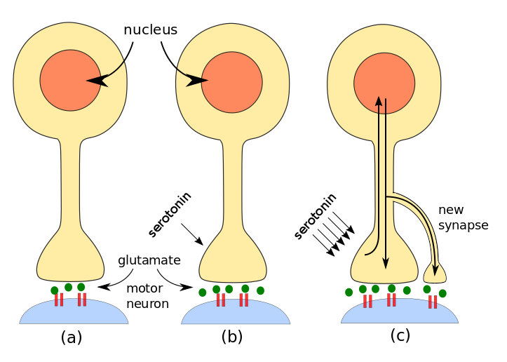
Steps in invertebrate learning: (a) simple stimulus, (b) short-term learning, (c) long-term learning. By author after Kandel.

Steps in invertebrate learning: (a) simple stimulus, (b) short-term learning, (c) long-term learning. By author after Kandel.

A series of shocks to the tail of Aplysia brings about long-term learning by the additional formation of more synapses. On the molecular level, the process begins as for short-term memory. A modulating neuron releases serotonin, but more of it than in the short-term case. Not only is PKA activated, but also a second kinase called MAP kinase.5Mitogen-activated protein. Both PKA and MAP move to the neuron’s nucleus in the cell body and activate a regulatory protein (transcription factor) called CREB.6Creb is cyclic AMP response element-binding protein. You see why it’s called CREB. There are two forms of CREB:

  • CREB-1 promotes gene expression and is activated by PKA;
  • CREB-2 inhibits gene expression and is activated by MAP.

So some genes are activated and some are suppressed in order for the appropriate proteins to be produced by the processes of transcription and translation already studied. The proteins and mRNA then travel back from the nucleus to the axon where they promote long-term synaptic facilitation through the growth of new synapses.  But this must only take place on the specific axon terminal, not others, and this requires yet another chemical substance.

A molecule called CPEB7CPEB is cytoplasmic polyadenylation element-binding protein. is found in neurons of Aplysia, Drosophila, mice and humans – and so probably every animal species. It is (or is similar to) a prion, i.e., it exists in two different conformations (shapes). Serotonin causes CPEB to switch from its inactive to its active form. Since the active form is present only in the axon terminal which has been modulated by serotonin, it can serve as a signal to the proteins and mRNA coming from the nucleus which axon terminals they should enhance by making new synapses. Very importantly, CPEB also assures the continuation of local protein synthesis using mRNA from the nucleus, in effect perpetuating the increased synaptic strength. This is long-term implicit learning.

It is also the only known case where the activated form of a prion serves a useful (to us) purpose. Other prions, such as those in mad-cow or Creutzfeldt-Jakob disease, are lethal.

What we have learned about implicit learning in invertebrates: Both short- and long-term memory depend on second messenger cAMP whose synthesis is controlled by serotonin. Whereas short-term implicit learning takes place locally at synapses, long-term memory requires contact with the nucleus and expression of new proteins in order to grow more synapses and reinforce short-term changes.

Learning in vertebrates

A neuron will fire an action potential if its total synaptic input is above a threshold. Normally, a single input will have little or no effect, but a number of inputs can have an effect. If

  1. an axon tries repeatedly to activate a cell which
  2. is already strongly activated by another or other inputs at the moment when the input under question arrives,

then the synapse of the first axon with the cell is enhanced in strength.

Similarly, but surprisingly, if the input arrives when the post-synaptic neuron is weakly activated, the synapse of the pre-synaptic neuron is diminished in strength.8The last two statements correspond to what is referred to as Hebb’s law.

Studies of London taxi drivers have shown clearly that the hippocampus is the seat of spatial memory, a type of explicit memory.  One way such synaptic strengthening can take place in the hippocampus is through the mechanism of long-term potentiation (LTP), which requires the synchronized firing of NMDA receptors and AMPA receptors on a post-synaptic terminal.9AMPA = α-amino-3-hydroxyl-5-methyl-4-isoxazole proprionate; NMDA = N-methyl-D-aspartate. Although both are glutamate-gated receptors, their function is somewhat different. AMPA receptors allow only Na+ and K+ to cross the cell membrane, whereas  NMDA receptors also allow large amounts of Ca++ to enter.

Normally, the channel of the NMDA receptor is blocked by the presence of a Mg++ ion, which keeps the passage of Ca++ minimal. Even if NMDA channels are gated by glutamate, they will remain blocked by the Mg++. Only if AMPA channels first adequately depolarize the target cell will the voltage-gated NMDA channels open. A low-frequency action potential in the pre-synaptic terminal releases only moderate amounts of glutamate, so the post-synaptic AMPA receptors allow in moderate amounts of Na+. However, a high-frequency pre-synaptic action potential releases much more glutamate which in turn causes the AMPA receptors to allow in much more Na+. Electrostatic forces due to the resulting increase in positively charged Na+ in the post-synaptic terminal push out the Mg++ from the NMDA receptors, which then allow large amounts of Na+ and, especially, Ca++ to enter the cell.10Why does the electrostatic potential push out Mg++ but allow Na+ to enter? This only happens if there is a significant amount of Na+ inside the cell and if the NMDA receptor has already bound glutamate outside the cell. So opening of the NMDA receptor requires both pre- and post-synaptic events, making it a coincidence detector and acting like an AND gate.

Once inside the cell, Ca++ acts as a second messenger and brings about a number of results. First, it causes AMPA channels already present in the interior of the cell to be inserted into the membrane of the receptor cell, thus strengthening the synapse. 

AMPA receptors recently installed during LTP disappear after a while if no other event takes place. But prolonged Ca++ presence can bring about production of cAMP and PKA which, as in implicit long-term memory, contribute to protein expression by an increase in the CREB transcription factors and synthesis of new AMPA receptors. They also lead to growth factors which can bring about new synapses between the pre-synaptic and post-synaptice neurons., again strengthening the connection.

One can see how this process can lead to associations in neuronal circuits. Suppose a neuron has synapses from three inputs, say, X, Y and Z. If X fires all alone, it is incapable of depolarizing the post-synaptic cell enough for LTP to occur. The same is true for each of the other two synapses. But if both X and Y fire simultaneously, i.e., repeatedly over the same short time period, their cumulative effect may be strong enough to depolarize the cell sufficiently for LTP to take place, meaning that the X and Y synapses will be strenghtened by the means we have seen. Now, if either X or Y brings an input, the post-synaptic neuron may fire, meaning that X or Y have the same result and are therefore associated. The third synapse, Z, will not have been enhanced.11This excellent example is based on one given by Bear et al.

Another possible means of synaptic enhancement may be due to the presence on dendrites of tiny “spines”, some of which have bulbous heads, which has led to their being described as shaped like mushrooms, doorknobs or even punching bags! They are thought to contribute to synaptic plasticity because they have both AMPA and NMDA receptors. They can appear and disappear, and grow and change their shape, depending on activity on both sides of the synapse. They also contain ribosomes, which are normally found near the cell body where they fabricate proteins from the recipes transcribed from DNA by mRNA. Their presence in dendritic spines is thought to be correlated with protein production which may also modulate synaptic strength. Research continues.

Other mechanisms, such as phosphorylation of AMPA receptors, are beyond the scope of this document.

A phenomenon similar to LTP, long-term synaptic depression, or LTD, in which AMPA receptors are removed from the synapse, explains the weakening of synapses in the case of a weakly activated synapse.

Another possible means of synaptic enhancement is due to the presence on dendrites of tiny “spines”, some of which have bulbous heads, which has led to their being described as shaped like mushrooms, doorknobs or even punching bags! They are thought to contribute to synaptic plasticity because they have both AMPA and NMDA receptors. They can appear and disappear, and grow and change their shape, depending on activity on both sides of the synapse. They also contain ribosomes, which are normally found near the cell body where they fabricate proteins from the recipes transcribed from DNA by mRNA. Their presence in dendritic spines is thought to be correlated with protein production which may also modulate synaptic strength. Research continues.

Other mechanisms, such as phosphorylation of AMPA receptors, are beyond the scope of this document.

Similar mechanisms must work during the modification of brain circuity during embryonic development.

We see now that Ca++ is extremely important to the organism for

  • formation of bones and teeth
  • neurotransmitter secretion in pre-synaptic cells
  • muscle contraction
  • neural plasticity.

Overview of senses and memory

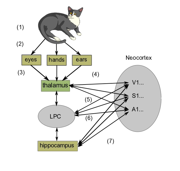
In brief, the learning process goes something like the following diagram, where we are learning what it is like to stroke a cat on our lap. The diagram shows only a schematic representation of the process, which is in reality much more detailed.

Schematic learning mechanism, by author

Schematic learning mechanism, by author

In order to see a greatly simplified version of what takes place in learning, we can regard it as taking place in three steps:

  • Perception
    1. First, we have the cat we are looking at, listening to (if it purrs) and stroking.
    2. Light and sound waves travel to eyes and ears, somatosensory perception takes place through the fingers.
    3. Signals produced in the sense organs in the retina (seeing the cat), hands (feeling its fur) and ears (hearing it purr) are transferred to the thalamus. (If the cat had just rolled around in something smelly, which well-brought-up cats do not do, olfactory information would go directly to the olfactory cortex before being projected back to the thalamus and then relayed to the cortex for consciousness.)
    4. The thalamus routes the information to the appropriate cortex areas (Visual, Somatosensory or Auditory Areas 1 and higher) for analysis. Many scientists think that the combined activity of these areas of cortex is what constitutes the perception of the experience
  • Formation of short-term memory
    1. Projections occur from the neocortex to the lateral prefrontal cortex (LPC), which creates temporary links to them. For our purposes, we may say this set of signals constitutes the image we “perceive” momentarily in our minds.
    2. The projections from the neocortex sensory areas to the LPC work in both directions. The LPC sends signals back to the cortex, bringing about activation of neurons similar to those evoked by the original image (sight + feel + sound). But this time, rather than originating in the sense organs, the signals are coming directly from the LPC. In this way, the LPC causes the set of neuron activations (the image) to remain in the neocortex longer than it might have done otherwise. This is working memory, one form of short-term memory. Most people are limited to no more than seven items at a time in working memory.12According to my wife, I am limited to only one – maybe two, if you include breathing.
  • Remembering
    1. The hippocampus also is connected to virtually the entire neocortex, as well as the LPC. Like the LPC, it can bring about a set of connections in the neocortex (i.e., alteration of synaptic strengths), which is at the core of thinking about the image in question. Repeated thinking like this is called rehearsal. Over time (maybe several days), such rehearsal, which takes place predominantly during REM sleep13Deep sleep in which dreaming is related to Rapid Eye Movement (REM)., leads to a long-term memory of the event, again, in the same part of the sensory cortex where the original sense-data experience was stored.

Consciousness and all that

To be quite frank, it seems that not a whole lot is really known about consciousness. It is the subject of much conjecture by both scientists and philosophers . Some one of these has suggested that consciousness may be considered as awareness expressed by language. Awareness is related to perception. Clearly, we need some more precise definitions of what all these things are.

We have skipped lots of subjects, perhaps more than in other chapters. Among further considerations which should be taken into account are the following:

  • Planning and decision making. Choice – do we have any?
  • Language and speech.
  • Sleep and waking.
  • Emotions, on which subject whole books have been written14Such as those of Antonio Damasio..
  • Repair and regeneration in the NS.

Notes

Notes
1 Edelman (2006).
2 I see it as the enhanced synapses’ having less resistance to passage of electrochemical neuronal circuits, so that signals take the path of least resistance from one neuron to another. Physicists like minimizing paths.
3 All the discussion of Aplysia is based on Kandel (2006).
4 We met cAMP when we looked at how the lac operon works.
5 Mitogen-activated protein.
6 Creb is cyclic AMP response element-binding protein. You see why it’s called CREB.
7 CPEB is cytoplasmic polyadenylation element-binding protein.
8 The last two statements correspond to what is referred to as Hebb’s law.
9 AMPA = α-amino-3-hydroxyl-5-methyl-4-isoxazole proprionate; NMDA = N-methyl-D-aspartate.
10 Why does the electrostatic potential push out Mg++ but allow Na+ to enter?
11 This excellent example is based on one given by Bear et al.
12 According to my wife, I am limited to only one – maybe two, if you include breathing.
13 Deep sleep in which dreaming is related to Rapid Eye Movement (REM).
14 Such as those of Antonio Damasio.

Motor output from the nervous system

We have already seen how afferent axons from sensory receptors enter the spinal column through the dorsal root and motor neurons exit through the ventral root. The afferent and efferent axons are grouped into spinal nerves before separating to go their separate ways. Once outside the spinal column, these axons and nerves are part of the peripheral nervous system, or PNS. We also have seen how efferent axons connect to muscle fibers via the neuromuscular junction (NMJ) to initiate muscle contraction. So not very much is left to add, unless we go into more detail.

As far as output from the nervous system is concerned, there are two parts:

  • the somatic motor system, which controls conscious perception and voluntary motor response, and
  • the autonomic nervous system (ANS), which is responsible for control, such as the glands, heart and involuntary muscles .

There are three sorts of movement, controlled by different neural circuits:

  • involuntary movements which regulate internal functions and homeostasis;
  • conscious voluntary movements;;
  • reflexive movements, automatic reactions which nevertheless may become conscious.

The third type, reflexive movement, is a fast, protective mechanism, which generally occurs before the brain is informed of the situation. Instead of the sensory neurons’ contacting the brain to obtain a command, these signals pass directly to interneurons in the spinal column and from there back out to the muscles, thereby avoiding the delay which would result from contacting the brain to obtain a response. This is of course how the classic knee-jerk reaction works. The interneurons are linked together into networks. The signal may also be passed to the brain to become conscious.

The anterior cingulate cortex (ACC) compares plans to how things are really happening in order to correct for errors or trouble due to unforeseen circumstances. It also monitors progress towards goals. It is connected to the lateral prefrontal cortex, giving it access to working memory.

We will not look any deeper  on the subject of motor control. So let’s go study how learning and memory take place.

Hearing, touch and taste

Hearing

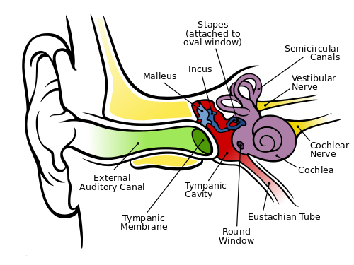
Hearing depends on ionotropic mechanoreceptors based on selectively depolarizing ion channels. First, sound energy is reflected and directed by the earlobe (pinna) , which can detect some vertical direction and modulates the frequency some as a function thereof. Then the energy passes through the auditory canal of the so-called outer ear, whose sensitivity is subject to resonance at the mid-tones we use to distinguish sounds. At the end of the canal, the sound wave hits the eardrum (tympanic membrane). This membrane is connected to the trio of tiny bones – the hammer (malleus), anvil (incus) and stirrup (stapes) – which use physical leverage to bring about slighter but stronger movement where the stirrup presses against the entry membrane to the cochlea, the oval window. The cochlea is rather like a nautilus shell, larger at the entry and spiraling somewhat to a smaller end. Its walls are lined with fine hairs which have finer hairs upon them, called cilia. As the cilia are moved by the sound energy, they stretch the hair-cell membrane, causing the stretch-sensitive mechanoreceptors in the membranes to depolarize. The resulting action potential sends information about the frequency and amplitude of the sound along the auditory nerve. The wider entrance of the cochlea detects higher frequencies, the small end, lower, so that frequency is a function of position in the cochlea.

The human ear, by Chitta L. Brockmann, via Wikimedia Commons

The human ear, by Chitta L. Brockmann, via Wikimedia Commons

Also visible in the figure are the three semicircular canals which constitute the vestibular system. They are oriented so that they can detect rotation and angular acceleration of the head about three axes and, so, in three dimensions. Not shown are the utricle and saccule, otolith organs which detect not only head tilts but the force of gravity. They and the semicircular canals are also lined with cilia and the set of vestibular and auditory cilia on either side of the head is said to constitute a vestibular labyrinth. Nerves from these systems are used by different parts of the brain in conjunction with ocular information and proprioception to detect and maintain the body’s orientation. They also contribute to the vestibulo-ocular reflex (VOR), which enables us to keep our eyes on a subject in spite of head movements.

The signals detected by the cilia are sent through other structures and finally arrive in the inferior colliculus of the thalamus on the opposite side of the brain. The thalamus relays the information on to the primary audio cortex, A1, in the superior temporal lobe, on whose surface location is a function of frequency, rather like the skin (somatosensory) map. It is not completely understood what happens after that. It is known though that two areas on the left side of the brain are important.

  • The zone called Wernicke’s area is necessary to the understanding of language.
  • Similarly, Broca’s area is necessary for producing speech.

Although these two areas are found on the left side of the brain in most right-handers, the right side also seems to be necessary for the comprehension of prosody (change of tonality and rhythm in speech). So it is only logical that the right side appears to play a role in the appreciation of music.

Detection of sound direction, though less good than in the case of vision, is accomplished vertically by the pinna, as already discussed, and horizontally by the difference in arrival time and intensity of the sound in the two ears.

Touch

Touch, or somatosensory perception, takes place in ionotropic receptors mostly in the dermis, the lower part of the skin. The outer part, or epidermis, is composed of sloughed-off dead cells from the dermis and constitutes a mechanical protection layer. The receptor cells themselves are in the dorsal root ganglion, a concentration of cells just posterior to the spinal cord. Such a cell has no dendrites and only one axon, which bifurcates, one end going to the dermis where its end holds the receptor, one into the spinal column. From there, it connects to interneurons which carry the signal up the spinal column to the thalamus on the opposite side of the brain from the receptors. In addition, the dermis contains free nerve endings with ion-channel receptors sensitive to temperature, extreme force or other possibly dangerous contacts.

The ventral posterior nucleus of the thalamus relays the signal to the somatosensory cortex (parietal lobe) just behind the central sulcus, where it is localized on a skin map, as already seen. In the map, the signals from a fairly well-defined part of the skin (such as the face or a hand) are found together. Outputs from the map connect to memories of previous touch-associated events. Cross-talk between adjacent areas of the map may lead to confusion as to the source of a stimulus.

Smell (olfaction)

The olfactory bulb in the top of the nose canal contains about 10 million olfactory receptors of about 1,000 types. They are metabotropic receptors on the cilia of the bulb. A smell is detected by many receptors and is distinguished by something like a weighted sum of the different results.

Olfaction is the only one of our senses whose receptors do not send information to the brain by way of the thalamus. Instead, they project to several sites in the cortex, including the olfactory cortex, which has direct influence on various brain areas. Information also passes via the olfactory tubercle to the thalamus, which transmits it to the orbitofrontal cortex in order to bring about consciousness of the odor. Other parts of the cortex control unconscious or reactive odor detection and memorization of smells.

The reason the olfactory receptors bypass the thalamus is evolutionary and is probably due to the fact that our ancestors already had a highly developed sense of smell, much like many animals today. So all that was left was to relay the smell to the thalamus in order to it to become conscious.

The receptors also send information to, among others, the amygdala for emotional response (e.g., for spoiled food) and to the hippocampus for memorizing of smells.

Taste

The surface of the tongue has structures called papillae, in which there are four types of receptors, only three of which are taste buds, containing about 10,000 taste cells. Each cell has several receptors responding to five different basic tastes: sweet, salt, bitter, sour (acid) and umami (meaty). Some are ionotropic, some metabotropic.

Signals pass from the receptors through another structure, the nucleus of the solitary tract (NST), to the thalamus either as pure basic tastes or mixtures. The thalamus relays it to the cortex for consciousness of the taste. Connections with the amygdala allow recognition of tastes associated with bad experiences in the past and so to be avoided. The NST, which influences the ANS, also receives input which may signal that you have eaten enough of that taste.

We distinguish different tastes far less well than different smells. Taste information is joined with olfactory information in the orbitofrontal cortex to generate flavor. Flavor therefore suffers if, for instance, our olfactory mucus is affected by a head cold.

Now, onward to brain output to the motor system.

What developmental biology tells us

What developmental biology tells us

Knowing about embryonic development is especially helpful in understanding the structure of the brain.

Fertilization, pre-embryonic development

Human pre-natal development traverses three stages:

  • The first two weeks of human development are called the pre-embryonic stage;
  • weeks 3-8, embryonic;
  • and after that, fetal.

Let’s start at the beginning.

Of the tens of millions of human sperm ejected during intercourse, millions are destroyed by the acid environment (pH 3.8) of the vagina or trapped by gluey cervical mucus. Thousands more are killed by uterine leukocytes.1Only strong sperm will make it. Force of numbers helps, too. The sperm follow the gradient of the chemical progesterone, which is released by the oocyte (egg). The surviving sperm usually meet the oocyte in the uterine (fallopian) tubes between the ovary and the uterus, since an unfertilized egg cannot live long enough to make the three-day journey along the tube. It is a question of force of numbers, as hundreds of sperm burrow through the oocyte’s outer protective cover, the corona radiata. Together, they release digestive enzymes from their tips to degrade both this layer and the inner layer, the zona pellucida. Finally, one sperm may manage to fuse its cell membrane with that of the oocyte so that it can release its DNA-containing nucleus into the oocyte. Then the work of the sperm is finished, as there exist mechanisms to prevent more than one sperm from fertilizing the oocyte. The process by which the haploid oocyte and the haploid sperm form a diploid zygote is rather more complicated than this, but that is the result.

Some time after fertilization, all the male mitochondria in the embryo are destroyed, leaving only the maternal mitochondria. It is not known how male mitochondria are recognized, but a gene has been identified, cps-6, which is imported into the male mitochondria and breaks them down. Since suppression of this gene leads to increased mortality in these embryos, there is certainly an evolutionary cause for the destruction, but it is not yet known.2How paternal mitochondria are destroyed in an embryo”, https://whyevolutionistrue.wordpress.com/2016/06/24/paternal-mitochondria-are-destroyed-in-an-embryo/.

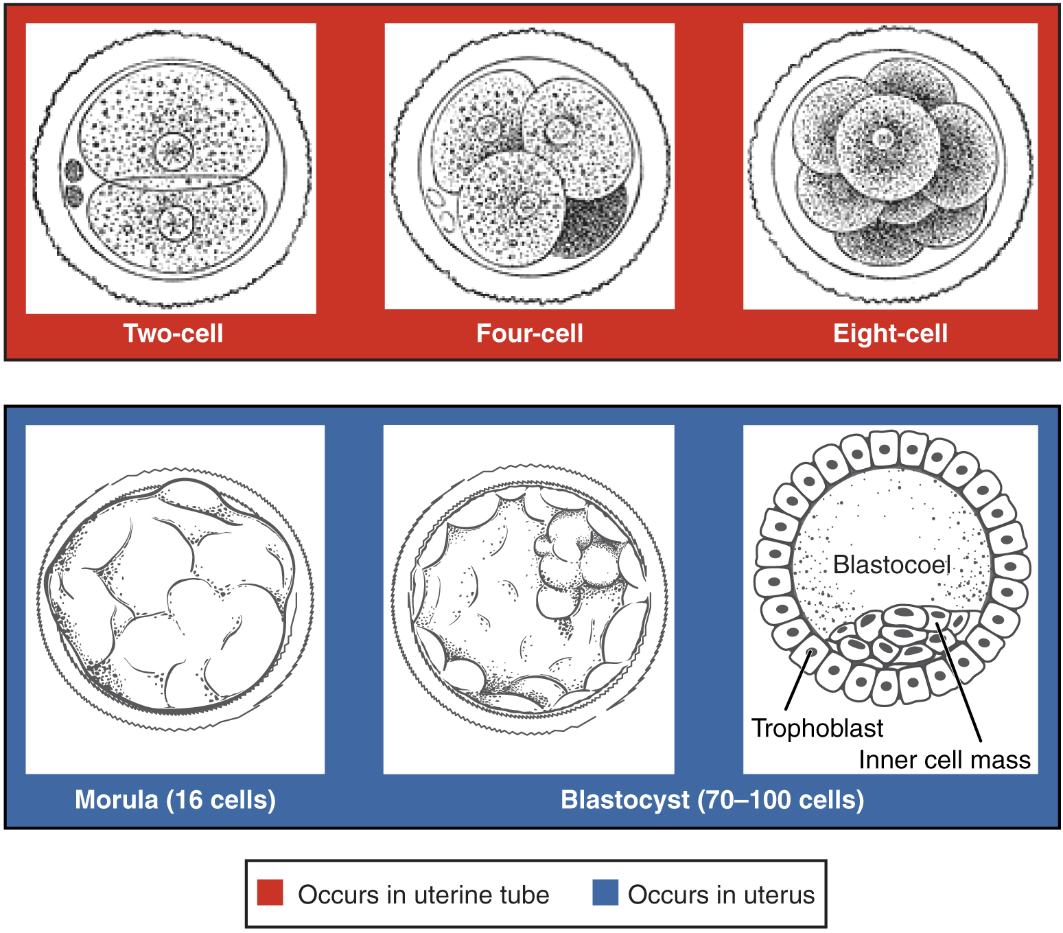
The conceptus, the zygote and its containing membranes, takes three days to reach the uterus. Along the way, mitotic cleavage increases the number of cells, called blastomeres, but without increasing the total overall size. By the time the conceptus reaches the uterus, it is composed of sixteen compacted cells, together called a morula. Mitosis continues, soon making about 100 cells arranged in a dense shell around a fluid-filled cavity called a blastocoel. The conceptus is now called a blastocyst. The cells of the outer shell are called trophoblasts and will form structures like the placenta.. Within the cavity, other cells form an inner cell mass which will become the embryo.3Information in these paragraphs on fertilization comes mainly from Wolpert (2011) and Openstax Anatomy and Physiology, http://cnx.org/contents/FPtK1zmh@8.25:FwQJfRAS@3/Embryonic-Development.

During this time, the inner mass is composed of totipotent, or pluripotent, stem cells, which can differentiate to form any type of cell.

Pre-embyronic cleavages, from Openstax College

Pre-embyronic cleavages, from Openstax College

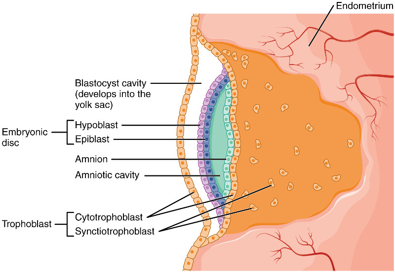
After a week, if all goes well, the blastocyst will attach to the inner uterine wall, the endometrium. In over half the cases, though, all does not go well and the blastocyst goes out with the menses. In successful implantation, the blastocyst eats through the endometrium, which then reforms so as to surround the embryo. The implantation marks the end of the pre-embryonic and the start of the embryonic state.

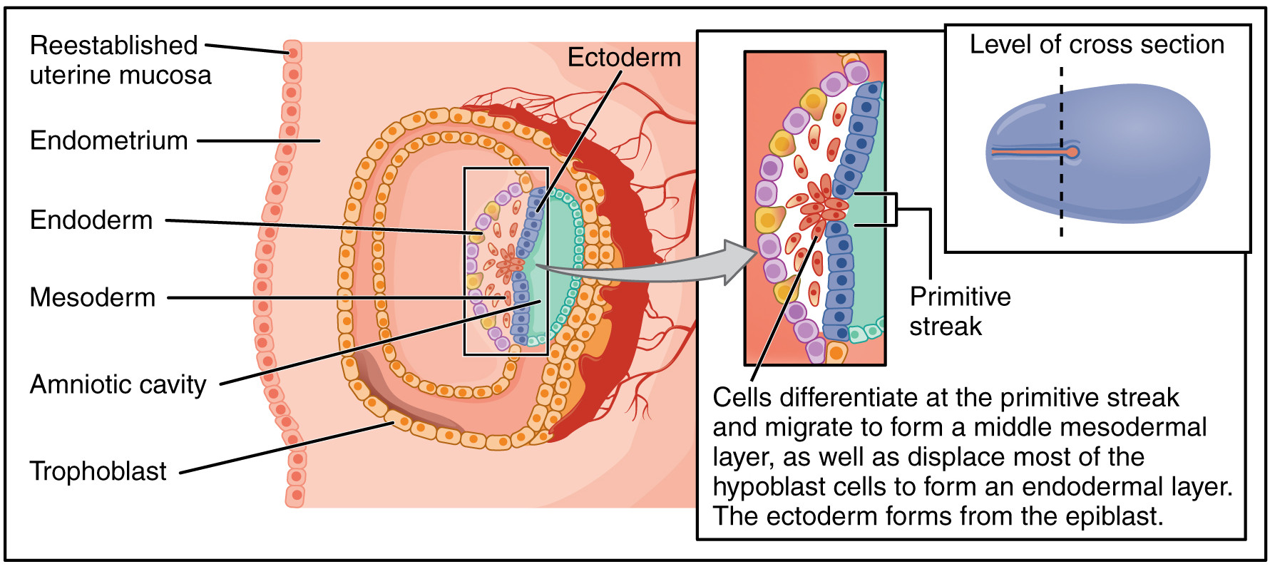
At the second week of embryonic development, the human zygote has developed into a two-layered embryonic disc contained between the blastocyst cavity (which will become the yolk sac) and the amniotic cavity. The ventral (forward) layer against the blastocyst cavity is the hypoblast. The dorsal layer against the amniotic cavity is the epiblast.

Human embryo before gastrulation, after Openstax College

Human embryo before gastrulation, after Openstax College

Gastrulation and embryogenesis – germ layer formation

At the third week, gastrulation takes place, converting the embryo from a two-dimensional disc into a complex three-dimensional structure formed of three germ layers. An indentation called the primitive streak forms along the dorsal (toward the back) surface of the epiblast. Growth factors produced by cells at the caudal (toward the posterior) end of the streak bring about cell multiplication. New cells migrate to the surface of the epiblast where they are specified as one of two new types, then migrate through the streak and form two new cell layers called germ layers.

  • One layer, called the endoderm, pushes aside the hypoblast and comes to lie against the yolk sac. The endoderm will form the epithelial lining of the internal organs, among other things.
  • A middle layer called the mesoderm is formed next to that. It will form the skeleton, muscles and connective tissues.
  • The remaining epiblast cells become the third germ layer, the ectoderm. It will form the integumentary (skin) and nervous systems.

In chordates (such as us), as the neural streak shortens and disappears, it leaves behind a rod-like structure called the notochord. The notochord will later become the nucleus pulposus, the viscous inner core of the inter-vertebral discs that can cause such excruciating back pain when they function badly.

The mesoderm along the notochord forms paired lateral bulges called somites. Positional identity along this axis is furnished by our old friends the hox genes. As we have seen, these genes are expressed in the same order in the DNA as along the antero-posterior axis of the developing embryo. From the somites will develop the spinal column and its 33 vertebrae, as well as associated skeletal muscle.

Gastrulation of the human embryo, after Openstax College

Gastrulation of the human embryo, after Openstax College

Three mechanisms are important for these changes:

  • Changes in cell shape are important in gastrulation and other processes. For instance, if a cell contracts on one side, it will become wedge-shaped. Joined to other cells with the same shape, an arc is produced, which is how a flat sheet of cells can bend. Such changes originate in the cytoskeleton.
  • Cells have proteins on their surfaces which bind selectively to proteins on other cells, thus causing specific cells to adhere together. Different adhesive molecules cause different cells to adhere to one another.
  • Cells can move relative to other cells by extending a long process called a filopodia, which is pushed out by elements of the cell cytoskeleton. The filopodia then move by a retraction mechanism similar to the one used by muscles to contract.

Neurulation – beginning of organogenesis and the nervous system

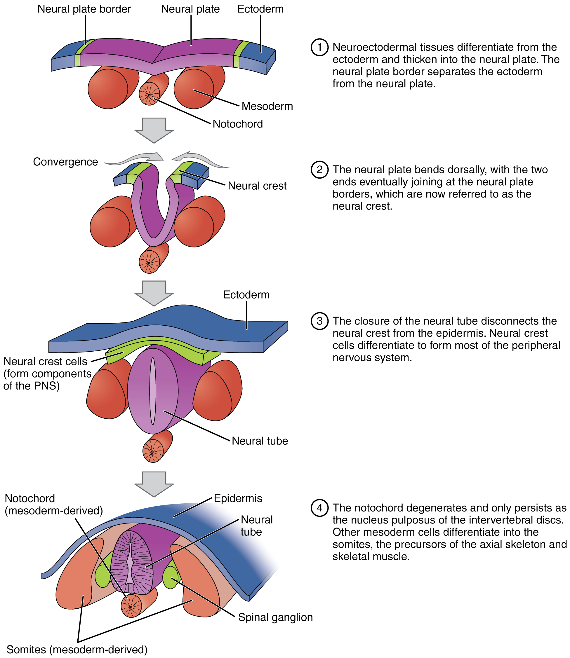
Soon after gastrulation, the rudiments of the CNS develop in the process of neurulation. Some cells of the ectoderm differentiate into neurepithelium and form a thickened area of the dorsal ectoderm called the neural plate. As shown in the figure, cells in the plate change shape and form a groove, the neural groove. The sides of the groove constitute the neural folds, which invaginate (grow out and come together to form a depression) and fuse, forming a neural tube. Cells along the tube change their adhesive properties so that the tube separates from the plate. The entire CNS will develop from the neural tube walls. The posterior part of the neural tube is formed differently, from a rod of cells which develop an interior cavity.

As the neural folds fuse, some ectoderm at the edges of the neural plate is squeezed off and is then situated between the neural tube and the ectoderm. This is called the neural crest and is the source of the entire PNS.

Neurulation forms the rudiments of the CNS and skeleton, from Openstax College

Neurulation forms the rudiments of the CNS and skeleton, from Openstax College

Organogenesis

By eight weeks after conception, the rudiments of all the basic organs have been set up. This is called organogenesis. Neurulation is the first important stage of organogenesis.

Germ_layers

Development of the germ layers in development, from Openstax College

The three germ layers develop into different tissues as shown in the figure. Not shown are the development of bones from the mesoderm, or the development of the CNS from the ectoderm.

The following table shows a brief outline of embryonic development.

Week Major events
4-5 The heart forms first as a tube, like the CNS, and starts beating around the beginning of week four. The liver starts to produce blood cells, a process which will later pass to bone marrow. Eye pits, limb buds and the beginning of the pulmonary system form.
6 Limb development starts in earnest.
7 Nostrils, outer ears and lenses form.
8 The major brain structures are in place. External genitalia are visible, but male and female are indistinguishable. Bone starts to take over from cartilage.

As it grows, the embryo folds so as to take on the familiar cylindrical shape, pointed at on end, with a primitive gut tube surrounded by the endoderm.

The organism is now at the stage of fetal development. From this point, we will restrict our interest to the consideration of nervous-system development.

Development of the nervous system

Some knowledge of the development of the nervous system is essential to understanding the structure of the brain.

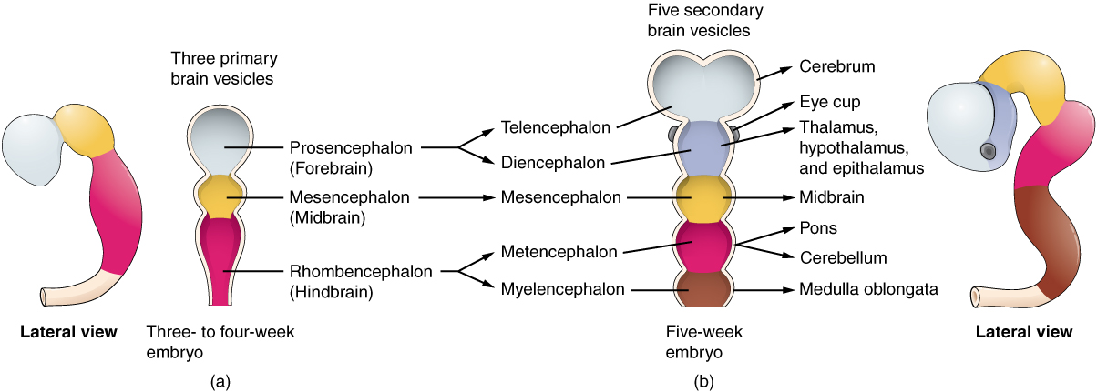
The anterior neural tube develops into the brain. It is first differentiated into three sac-like primary vesicules, from front to back:

  • the prosencephalon (forebrain)
  • the mesencephalon (midbrain)
  • the rhombencephalon (hindbrain), connected to the caudal neural tube.

As the prosencephalon begins to differentiate, two pairs of secondary vesicles grow on either side:

  • the optic vesicles, which will eventually become the optic nerves and the retinas (so the retina is part of the brain); and
  • the telencephalic vesicles, or telencephalon.

The remaining unpaired part of the prosencephalon is called the diencephalon, or “between brain”. The telencephalic vesicles grow out, then bend back over the diencephalon. From them grow another pair of vesicles, the olfactory bulbs. Cells within the telencephalon develop, forming new structures and axon systems. Spaces remain between the sides of the telencephalon and the diencephalon; they will fill with cerebrospinal fluid (CSF) and become the ventricles of the brain. The neurons within the gray matter of the telencephalon form the cerebral cortex and the basal telencephalon. The diencephalon differentiates into the thalamus and the hypothalamus. Axons from the developing forebrain extend to connect with other parts of the nervous system.4This brief summary is based on Bear, 178-192.

Brain vesicle development, from Openstax College

Brain vesicle development, from Openstax College

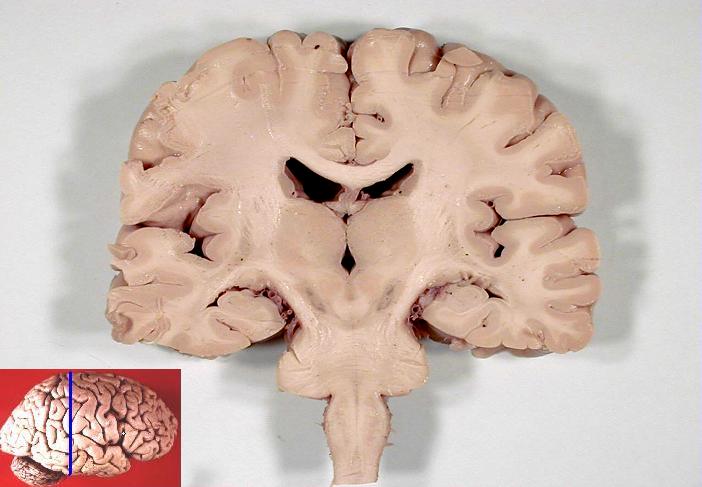
The hindbrain develops into the pons, medulla oblongata and cerebellum. In a coronal (side to side) section of the brain, it is easy to see how the telencephalon has grown down laterally to the diencephalon. The ventricles are also visible as dark areas.

Coronal section of human brain, from John A Beal, via Wikimedia Commons

Coronal section of human brain5Looks a bit like the Hulk, doesn’t it?, from John A Beal, via Wikimedia Commons

The following diagram shows a somewhat more complete schema for the development of the human brain. The examples of development of germ layers are quite incomplete.

Schematic diagram of human brain development, by author after Bear et al.

Schematic diagram of human brain development, by author after Bear et al.

Next, let’s go to the next subject, the central and peripheral nervous systems.

Notes

Notes
1 Only strong sperm will make it. Force of numbers helps, too.
2 How paternal mitochondria are destroyed in an embryo”, https://whyevolutionistrue.wordpress.com/2016/06/24/paternal-mitochondria-are-destroyed-in-an-embryo/.
3 Information in these paragraphs on fertilization comes mainly from Wolpert (2011) and Openstax Anatomy and Physiology, http://cnx.org/contents/FPtK1zmh@8.25:FwQJfRAS@3/Embryonic-Development.
4 This brief summary is based on Bear, 178-192.
5 Looks a bit like the Hulk, doesn’t it?

The lymphatic system

The lymphatic system, constituted of the lymph vessels and nodes and the lymph, serves several functions.

A partial, parallel venous system

In the circulatory system, the heart pumps blood throughout the body via arteries, then smaller arterioles and finally through tiny capillaries, from which molecules are exchanged with cells and the rest of the body. Due to the pressure of the blood, much plasma is also released. The endothelial walls of the capillaries are just one cell thick and the spaces between them allow fluid to pass, but not the larger erythrocytes (RBCs), which therefore remain in the blood.

Outside the capillaries, the escaped fluid and molecules constitute the extracellular or interstitial fluid. It is composed of WBCs, including lymphocytes, some hormones, glucose and other molecules, such as proteins and lipids. Much of it is reabsorbed, but some remains outside. One of the jobs of the lymphatic system is to carry it back into the blood stream.

The lymphatic system therefore can be considered a parallel path to the venous system, but it carries no RBCs, only WBCs, called leukocytes. The lymph, as the fluid is called when it is inside the lymphatic system, is returned to the veins at two ducts placed strategically where blood pressure is relatively very low and the liquid can therefore be inserted into the vein easily.

Lymph is not pumped by the heart. Smooth muscles in the walls of the lymph vessels, as well as skeletal muscle, squeeze the lymphatic vessels, pushing the lymph through one-way valves which prevent its returning to the capillaries. Similar valves allow the interstitial fluid to enter the lymph vessels but not to leave them. Lymph flows from lymphatic capillaries, or terminal lymphatics, and into bigger lymph vessels before re-entering blood veins in either the right lymphatic duct (which drains the upper right-hand side of the body) or the thoracic duct (which drains all the rest).

Immune system function

The lymphatic system is much more than a simple alternate return route for WBCs. Since it carries lymphocytes, it is crucial for the immune system. Scattered about the body along the lymphatic vessels are some 500-600 special tissues (too small to be called organs) called lymph nodes, in which immune-system cells do much of their work.

There are macrophages present in all tissue, but most of the immune-system cells spend their off-duty hours in the lymph nodes. The complicated process of B-cell and T-cell growth and activation (to be discussed in the next section) is carried out more rapidly in an environment where macrophages or dendritic cells and B and T cells proliferate and this is in the lymph nodes. The large number of such cells, as well as macrophages and others, means that the lymph passing through the node has most if not all its pathogens filtered out and phagocytozed.

Lymphatic vessels, from Openstax College

Lymphatic vessels, from Openstax College

The spleen is not connected to the lymphatic vessels, but It contains a large number of macrophages and dendritic cells which carry out a similar filtering process not on lymph, but on the blood.

Other secondary lymphoid tissues are:

  • lymphoid nodules, such as the tonsils, in the respiratory and digestive tracts, which contain dense clusters of lymphocytes;
  • mucosa-associated lymphoid tissue (MALT);
  • bronchus-associated lymphoid tissue (BALT).

Function with other molecules

Glucose in the small intestine diffuses out of the intestine, into capillaries and thence throughout the the body through the circulatory system. But lipids leave the small intestine in the form of chylomicrons, which are too large to enter capillaries. However, they can enter the lymphatic system lacteals, lymphatic capillaries in the intestinal villi. The lymphatic system then conveys the lipid-bearing chylomicrons into the blood.

In a similar way, other molecules, such as some hormones, or waste products, being sent to the liver or kidneys, and which are produced not in the blood but elsewhere in the body, may not be able to enter blood capillaries, and so flow into the circulatory system via the lymphatic system.

Now we go on to the immune system.

The immune system

The immune system is complex, multi-leveled and multi-component.1Its complexity and numerous components, which are not independent or separable for study, has led to confusion in the designation of its sub-systems. (Author’s opinion.) Even though much has been learned about it in recent decades, it is still under intense investigation.

Rather than starting out with lists of the components, the structures and so forth, let’s look at an example.

Overview – physical barriers and inflammation

Suppose something bad – a virus or a mean bacteria – gets into a body. What happens then to protect against this pathogen?

The first line of defense against pathogens is largely mechanical or chemical. It comprises such things as the skin, which forms a natural barrier to keep things out; the mucus in our respiratory system, which captures microbes in the air we breathe and, by means of tiny hairs – cilia – drags them back up to where we can swallow them2Yuk.; and the gastrointestinal system, which is highly acid and can therefore kill many invaders.

But some of the invaders get past these barriers. And some do not even need to, because they show up internally. And tissue damage can result from external sources also, as when a hammer is injudiciously applied to a thumb. So after the barrier layer, what then?

The next step is often inflammation, called the inflammatory response. The infected area turns red, swells up, becomes hot and hurts. This region is the battlefield where the fight carries on after the barriers have been breached. This is where the various cells of the immune system go about protecting us.

Injured cells call for help by releasing chemical messengers called chemokines into the interstitial fluid. These indicate to cells farther away that there is a problem to be solved and implores their assistance. The cavalry arrives not by following a trumpet call, but by moving against the gradient of the chemokines, toward their point of greatest concentration, in a process called chemotaxis.3Neutrophil to driver: “Follow that pathogen!”

Among those coming to help, mast cells are one of a group of cells which contain granules, from which they release histamine, leukotrienes and prostaglandins. These chemicals cause vasodilation: Blood vessels expand so more blood can come in, bringing help, but they also become porous. Increased blood flow and plasma leakage into the interstitial space are responsible for the heat and redness and painful swelling. 

Chemokines also lure in phagocytes to surround and absorb pathogens. The first ones to arrive are neutrophils, the most plentiful phagocyte in the body. Other phagocytes — macrophages and dendritic cells — come to phagocytize pathogens,  to engulf them and destroy them in the process of phagocytosis.. All the cells mentioned so far are part of the non-specific or innate response. They are non-specific because each of them may act against any pathogen. Because they all rush in, they operate quickly, but they are neither very precise nor efficient.

Now we reach the next level of the immune system, the specific immune system, or adaptive immune response, as B cells and T cells begin to do their thing.

Any invading organism consists of at least one compound — in it or on it — which can be used to identify it uniquely. These compounds are called antigens, from antibody generations. The adaptive response depends on these markers to distinguish one pathogen from another. Phagocytes, like macrophages. dendritic cells or B cells, extract the antigens from the pathogen itself and display them on their own surface. This enables a specific response, one depending on the identity of the pathogen. Such a series of events takes more time than the innate response, but because it is specific to the particular pathogen, it is usually more effective.

Macrophages which display antigens on their surface serve as messengers or links between the innate and adaptive responses and begin the intricate process by which B cells, with the collaboration of T cells, produce antibodies, which can protect us over time.

The complement system is composed of proteins which aid immune response. They may, for instance, bind to a pathogen and thus label it – opsonization – calling attention to it so that phagocytes will deal with it. The complement system can help out both the non-specific and specific responses.

Now let’s look at these different immune-system components in more detail.

Innate immune response

The innate, or non-specific, immune system depends on several types of cells. It most likely evolved before the adaptive immune response.

One group of cells is constituted of phagocytes, which can engulf and contain pathogen cells, a process called phagocytosis. The contained pathogen, called a phagosome, is usually killed by a lysosome in the phagocyte cell. A phagocyte recognizes pathogens in a somewhat approximate way, as its surface (not to mention its genes) can contain only a certain number of antigen pattern recognition receptors (PRRs). Phagocytes are also important because they represent the first step in the process which identifies pathogens and leads to the release of specific antibodies. There are several phagocytes associated with the innate immune response.

  • A macrophage is an amoeba-like phagocyte which can squeeze through tissues and capillary walls. It is capable of phagocytozing quite large bacteria.
  • A neutrophil is a spherical phagocyte summoned from the blood stream. It is a granulocyte, meaning that it contains granules of substances such as histamine. It is the most abundant of the white blood cells (WBCs).
  • A dendritic cell is an irregularly shaped cell with many appendages looking like the dendrites on neurons, hence the name. However, they are not neurons. Although they may be partly phagocytotic, their principal function is as antigen-presenting cells, about which more very soon.
  • A monocyte is a precursor cell which may mature into either a macrophage or a dendritic cell.

A natural killer cell (NK) is not generally cosidered a phagocyte, but it is capable of convincing a pathogen cell to commit suicide, or apoptosis. It may do this through the use of a special ligand or by releasing perforin to bore a hole in the cell wall and then cause granzyme to enter the cell and bring about apoptosis.

Antigen-presenting cells (APCs)

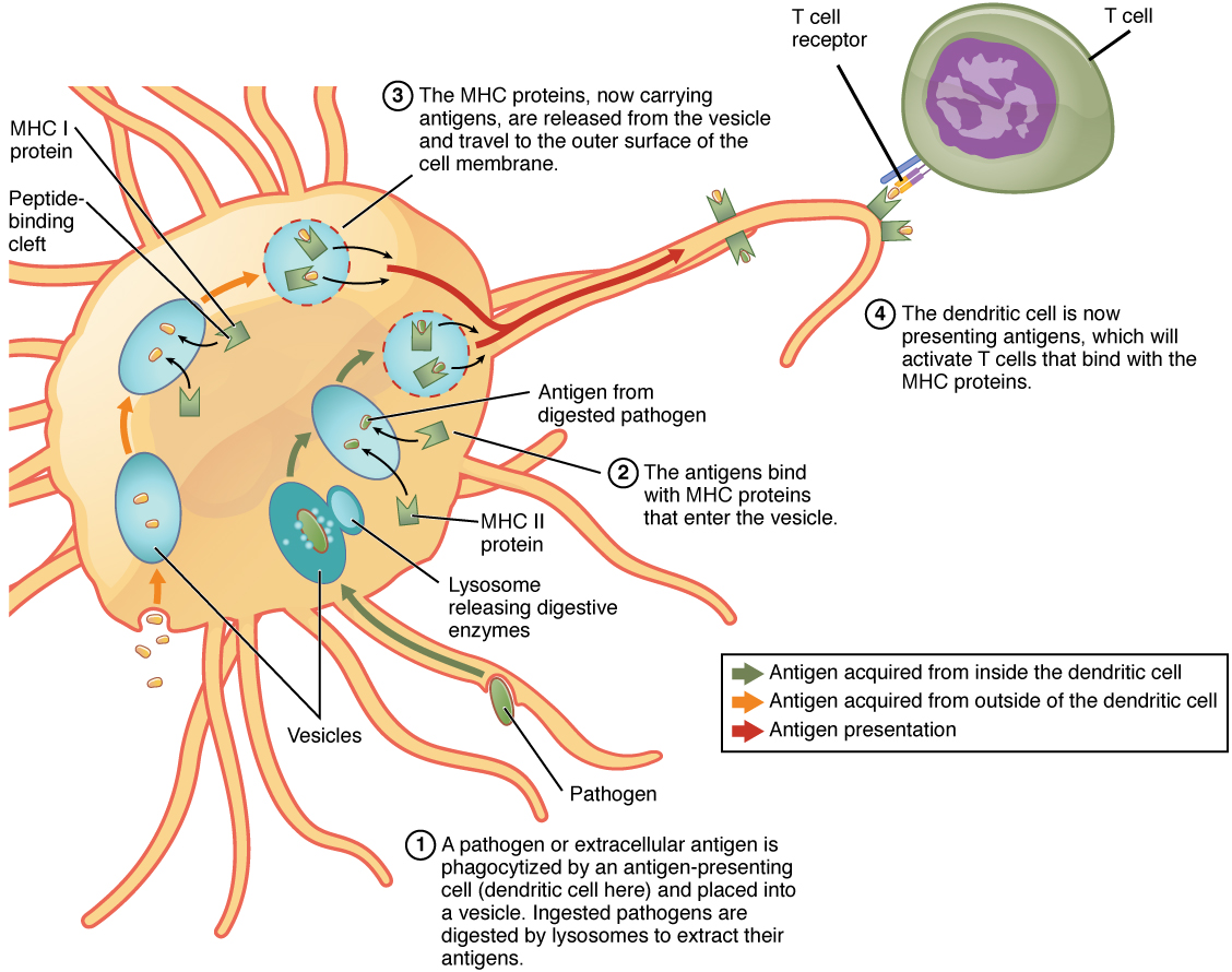
The mechanisms of antigen presentation are somewhat complex. T cells, an essential part of the adaptive immune system, recognize antigens only when they are presented on the surface of antigen-presenting cells (APCs). APCs internalize the pathogen or antigen, break It up and bind pieces of it with a protein called the major histocompatibility complex (MHC) molecule. Only when the complex formed by the MHC molecule and the antigen is presented on the surface of the APC can it be recognized by T cells.

There are two types of MHCs and so two types of APCs.

  • The so-called professional APCs are the innate-response macrophages and dendritic cells as well as the adaptive-response B cells, all of them cells of the immune system. When such a cell phagocytizes an external pathogen, it associates pieces of the antigens with MHC class II (MHC II) molecules and displays the complex on its surface. 
  • Non-professional APCs include all nucleated cells in the body.4Reminder: red blood cells lose their nuclei. They are not phagocytes, but they nevertheless display class I MHC (MHC I) complexes of internal pathogens, such as viruses which have reproduced within the cell.

Professional APCs can display MHCs of both classes, depending on whether the pathogen is internal (MHC I) or external (MHC II).

Antigen presentation, from Openstax College

Antigen presentation, from Openstax College

Leukocytes, lymphocytes and WBCs

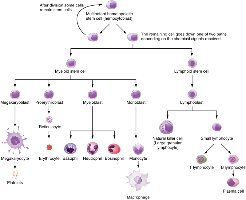
Blood cells are all differentiated from hematopoietic stem cells. For granulocytes like neutrophils, this occurs only in the marrow of bones . But lymphocytes and plasma cells are produced in lymphogenous material, which exists in lymph glands and other areas, including bone marrow

Differentiation of hematopoietic stem cells, from Openstax College

Differentiation of hematopoietic stem cells, from Openstax College

Erythrocytes are red-blood cells, of which the adults contain no nuclei, and platelets are responsible for blood clotting. The other cells shown are all WBCs, or leukocytes. In the lymphatic system, they are called lymph. They also constitute the interstitial fluid between cells.

Dendritic cells are special, existing in several varieties, and can come from a myeloid or a lymphoid precursor, not shown in the figure.

NK cells, B cells and T cells are called lymphocytes. Whereas the NK and B cells mature in bone marrow, T cells migrate to the thymus for maturation. NK cells are part of the innate immune system. B and T cells are cells of the adaptive immune system.

Each instance of a B or T cell has surface receptors which can be activated by only one type of antigen. Therefore, in order to detect almost any pathogen, B‑cell and T‑ell receptors exist in souch a great number of variiants that it would be impossible for so many genes to exist in their DNA. This difficulty is overcome by means of an astounding technique – gene shuffling. During the expression of their genes, segments of genes are shuffled like cards, giving over 1011 possible combinations, each of which detects a specific antigen.5Isn’t this amazing?

After strong selection against cells which may attack their own organisms, both types of cells migrate to lymphoid tissue throughout the body.

When a macrophage phagocytizes a pathogen, it passes antigens on to the lymphocytes. When a lymphocyte receives its specific antigen, it becomes activated and reproduces abundantly. (B cells also require the help of special T cells, as we shall see in a moment.) Activated B cells produce antibodies. Groups of antibodies or T cells activated by and sensitive to a specific pathogen are called clones.6A clone is not a simple copy, it is a set of copies sensitive to a specific antigen. Cells of a clone therefore share the same antigen receptors. Only lymphocytes which are activated by an antigen reproduce and multiply into clones, so the process can be seen as one of natural selection. Only activated cells survive and reproduce.

Adaptive immunity

Now for more details. I find this subject easier to understand by proceeding from less complex to more, so I will start with B cells, which will leave them partly unexplained until T cells have been discussed.

Humoral response – B cells and antibodies

When a “naive” B cell leaves the bone marrow or the lymphatic system and binds to an antigen, its ctivatiion process has begun. Part of the antigen is internalized, broken up and displayed on the cell’s surface in an MHC II complex. When this is bound by a type of T cell called a Th2 cell (the part that is left until later, but not much), the B cell is completely activated. (Some B cells are T-cell independent and do not require such activation.) The B cell then is duplicated into sets of clones to make many B cells of two types – effector cells and memory cells. The effector cells, now called plasma cells (not to be confused with blood plasma, which is mostly water) release a form of their surface receptors called antibodies, or immunoglobins (abbreviation Ig), into the environment.

The immunity conferred by antibodies is called humoral immunity. Each antibody recognizes only the antigen specific to its parent B cell. When antibodies – immunoglobins – encounter a pathogen carrying this antigen, they bind tightly to it via a lock-and-key mechanism based on their respective shapes and thus prevent the pathogen from doing any harm. They also signal phagocytes to absorb and kill the pathogen (opsonization again).

The memory cells wait around until they die or are needed again to clone and produce more receptor and memory cells. Some of them remain available for years or even decades. When needed, they allow for a much more rapid response to subsequent infections, called secondary response, as opposed to the initial, primary response. In secondary response, B cells as plasma cells are already available for producing antibodies without going through all the rigmarole of APCs and being activated by a T cell. We will look at why this takes place later.

Antibodies exist in five classes, imaginatively called, IgA, IgD, IgE, IgG and IgM.

  • Only IgM and IgD function as receptors on a B cell; IgD remain on the B cells.
  • IgM can leave the B-cell surface. They are the largest of the Igs, having 10 binding sites, whereas IgA has four and the others only two. So IgM is an excellent binder, especially during the early part of a primary response. In the latter part of the primary response, IgM can undergo a process of class switching in which it changes to a class IgG, IgA or IgE.
  • IgG is the most common (80% of antibodies in serum) and is the most important antibody in secondary response. It also is the only one which can cross the placenta to protect the fetus.
  • IgA has two forms. The eight-chain structure moves into mucous membranes and so is the only antibody to leave the interior of the body. The four-chain form remains in the blood.
  • IgE is very rare. It serves to make mast-cell degranulation specific. In doing so, it contributes to allergies.

Cell-mediated response – T cells

Like B cells, T cells exist in some 1011 possible versions, each receptive to a specific antigen. But T cells are more complex (which is why we left them until now), being of two types, one of which has sub-types.

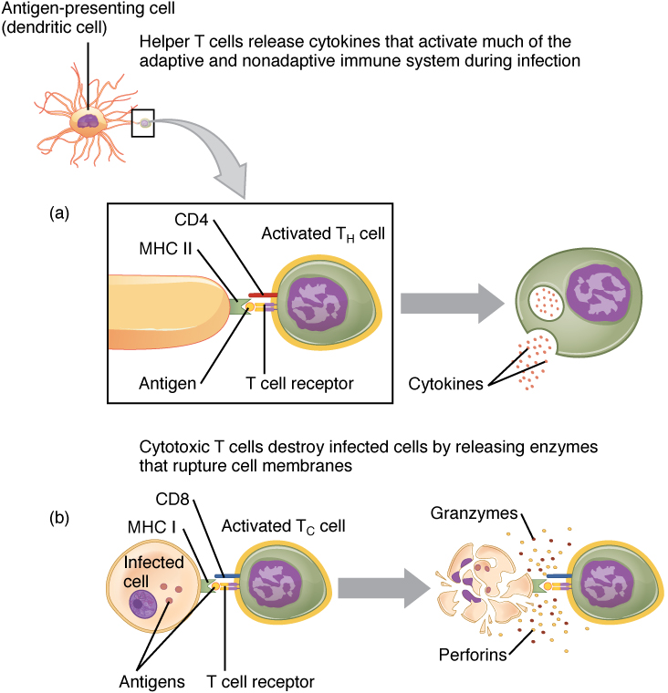
T cells can not detect antigens directly; they can only detect those which are displayed on the surface of APCs. Just as there are two types of APCs, according to their MHC type, there are two types of T cells. Certain glycoproteins on T cells are called clusters of differentiation, or CDs. On leaving the thymus, most T cells have either CD4 or CD8 and so are called CD4+ or CD8+.

  • CD4 is a co-receptor which enables so-called helper T cells, or Th cells, to recognize class II MHCs on APCs, including those on B cells.
  • CD8 is specific to MHC I and to cytotoxic T cells, also called Tc cells.
  • There is a third type, called Treg, for regulatory T cell, with CD4 and CD25.

Tregs are less well understood, but suppress immune response by other T cells in certain cases.

T-cell activation by APCs, from Openstax College

T-cell activation by APCs, from Openstax College

Of the two principal types, those simpler to understand are the CD8+ cells, called cytotoxic T cell or Tc cells when they are activated. As in the case of B cells, activated T cells multiply into sets called clones and while some of these become memory T cells, awaiting the next infection, the others become effector T cells. Effector T cells destroy infected cells by inducing apoptosis, like NK cells.

The other main type of T cell is the helper T cell, or Th, which exists in two versions, Th1 and Th2, the difference being mainly in the type of cytokines they secrete. Cytokines are short-distance signaling molecules. Those secreted by Th1 act as an alarm signal and promote phagocytosis. Alas, helper T cells are damaged by the human immunodeficiency virus (HIV).

B cell activation, from Openstax College

B cell activation, from Openstax College

Th2 is the one we have been putting off and which allows the B cell to do its job of distributing antibodies. In order to have any effect, a Th2 cell must recognize its own antigen type on a B cell’s class II MHC. When this happens, the Th2 cell secretes cytokines which, when detected by the B cell, complete activation of the B cell, so that it may clone to memory cells and antibody-emitting effector cells. A possible reason for the complexity of this procedure will be explained shortly.

Quick (?) overview

Let’s do a run-over of the humoral immune system, the creation of antibodies7Remember, we use the words immunoglobin and antibody to refer to the same things.:

  • A phagocytic cell of the innate immune system, such as a dendritic cell, phagocytozes a pathogen. It presents antigens from the pathogen on its surface in MHC II complexes.
  • A B cell of the adaptive immune system whose membrane-bound immunoglobins are of a specific type happens to meet the same type of pathogen and also phagocytozes it and presents its antigens on its surface in MHC II complexes.
  • Both the dendritic cell and the B cell are now APCs for the specific antigen. These two steps could have occurred in either order.
  • A helper T cell for the same type of antigen bumps into our dendritic cell, binds to the MHC II complex and becomes activated.
  • If the activated cell is a type Th1, it starts emitting cytokines as an alarm and to promote phagocytosis.
  • If the activated cell is a type Th2, it may later meet our B cell and bind to its MHC II complex. It then release cytokines which complete activation of the B cell.
  • The activated B cell duplicates itself into clones of two types, effectors and memory cells, the former being plasma cells which emit numerous immunoglobins – antibodies.
  • The antibodies cling to their specific pathogens and render them harmless. At the same time, they tag them for phagocytosis.

That was the case for an external pathogen. But some pathogens, like cancers, may occur internally or, like viruses, penetrate into the cell before being noticed. Since all nucleated cells may present MHC I complexes, these will be recognized by CD8+ cells which are activated to cytotoxic T cells which will act to kill the offending (or offended) cell.

Once activated, all B and T cells duplicate to form clones of numerous effector cells and memory cells.

Why so complicated? – auto-immunity

Why do B cells have to go through a process of being activated by T cells (which have to be activated by APCs…) before producing antibodies in the primary response? One answer is to avoid auto-immunity.

Auto-immunity is when your immune system mistakes some essential part of you for a pathogen. This can lead to some really awful conditions like AIDS, lupus or myasthenia gravis. Since the receptors on B and T cells are the result of gene shuffling, many combinations will occur which could lead to auto-immunity. This is avoided in two ways:

  1. Under the assumption that pathogens are rare within the bone marrow and the thymus, any B or T cells which activate inside them are killed before they can get out and do any harm.
  2. The probability that either a B or T cell released into the body be an auto-immune cell is small (because of step 1), The process of requiring that a T cell “vet” the activation of a B cell through the APC process means that the total probability of generating an auto-immune cell is something like the product of the individual probabilities and therefore very low indeed.

So the requirement that B cells be activated by their corresponding activated T cell is a way of avoiding auto-immunity. Nevertheless, however low the probability, some auto-immune diseases do occur … alas.

Now on to embryonic development, how that sperm and egg made us.

Notes

Notes
1 Its complexity and numerous components, which are not independent or separable for study, has led to confusion in the designation of its sub-systems. (Author’s opinion.)
2 Yuk.
3 Neutrophil to driver: “Follow that pathogen!”
4 Reminder: red blood cells lose their nuclei.
5 Isn’t this amazing?
6 A clone is not a simple copy, it is a set of copies sensitive to a specific antigen.
7 Remember, we use the words immunoglobin and antibody to refer to the same things.

The human circulatory system

We will consider the circulatory system only from a relatively high-level (undetailed) view of its structure and function – as a kind of plumbing system. This consists of several parts – the pump (the heart), the pipes (the veins and arteries) and the fluid (the blood).

The heart as pump – circulation

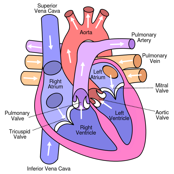
The heart consists of four chambers, two smaller, upper chambers – the atria (singular atrium) – and two larger, lower ones – the ventricles.

Structure of the human heart, from Wikimedia Commons

Structure of the human heart, from Wikimedia Commons

Even though we already indicated in the chapter on bioenergetics the general flow of blood through the heart and the cardiovascular circulatory system. it is worth repeating.

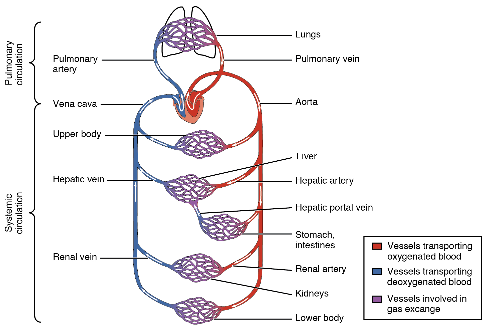
Oxygen-depleted blood from the body enters the right atrium of the heart and is pumped into the right ventricle and thence through the pulmonary artery into the lungs. (The figure is somewhat confusing because the lungs are shown above the heart, about at the place of the brain.) After picking up oxygen in the arterioles of the lungs, the blood returns through the pulmonary vein into the left atrium of the heart. It then is pumped into the left ventricle and out into the body. In capillaries, oxygen passes into cells and the oxygen-depleted blood is then returned to the right atrium through veins and either the superior or inferior vena cava.1Remember, veins flow towards the heart, whether they contain oxygenated blood or not. Four valves – the mitral, tricuspid, pulmonary and aortic – keep the blood from flowing back in the wrong direction.

Cardiovascular circulation, from Openstax College

Cardiovascular circulation, from Openstax College

The walls of the heart are composed of three layers of tissue, of which the most important is the myocardium. This is the muscle which contracts to squeeze the four chambers and force blood through the heart and out into the lungs or body. Being mostly muscle, the heart itself requires energy and nutrients and, so, blood. This is supplied by the coronary arteries branching off the aorta, the main exit for oxygenated blood. The heart pumps blood to itself.

Electricity in the heart

Where study of the heart becomes really interesting is its electrical operation, where we meet our old friends energy and communications again.

The big question is, “What makes the heart beat?” Surprise: The commands to make it do so do not come from the brain. The brain can only modulate the rate of heartbeat.

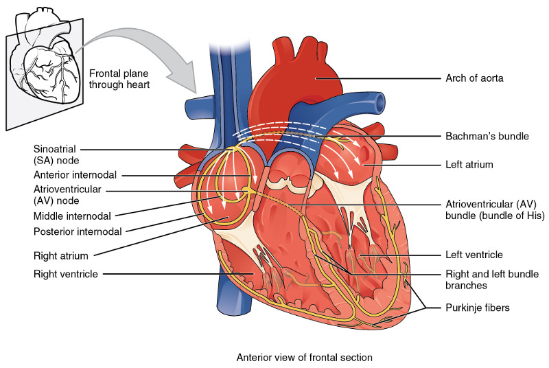
The generation of an electrical signal to make the heart beat, i.e., to cause its muscles to contract in a rhythmic and well-synchronized manner, takes place in the sinoatrial (SA) node (or SAN, or sinus node) in the wall of the right atrium near the entrance of the superior vena cava. (See following figure.) Although similar signals may be generated elsewhere in the heart, they are usually overwhelmed by the higher-frequency SA signal. Like most cells in the body, the cell membranes of heart muscle contain a number of structures for allowing – or requiring – substances to enter or leave the cell. (We considered this in the biochemistry chapter.) Among these are Na/K pumps which maintain a negative voltage inside cell walls. In the SA node, however, instead of a voltage of around -80 Mv, slow leakage limits the voltage to -55-60 mV.

Conduction system of the heart, from Openstax College

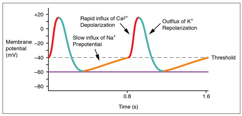
Heart beat is caused by the generation of an action potential in the SAN.

The initial increase of the SAN membrane potential from its minimum, rest potential, is due mostly to leakage channels which allow some Na+ to enter the heart, gradually raising the membrane potential from about -60 mV to -40 mV (the “prepotential” in the figure). This is the threshold value for a gated channel to open, allowing depolarization of the cell. However, because of the leakage, only slow Na+–Ca++ channels are opened, which explains the relatively slow rise of the potential. (The slow rate of potential rise from -40 mv to 10 mv is not obvious in the figure.)

Action potential at the SA node, from Openstax College

After about 100-150 milliseconds, the Na+–Ca++ channels close and K+ channels open. As K+ rushes out of the cell, the potential redescends to a bit under its initial resting value. Then the K+ channels also close and the cycle starts over. The result is that the heart beats all by itself due to the membrane potential of the cardiac muscle cells, the slow leakage of Na+ and the voltage-gated Na+–Ca++ and K+ channels.

The action potential generated in the SAN passes into atrial muscle fibers which contract and squeeze the atria. The action potential travels through the atria and down to the atrioventricular node (AV node), near the floor of the right atrium. A delay takes place here which keeps the ventricles from contracting at the same time as the atria. The action potential then passes into the bundle of His, which separates into two different paths, the left and right bundle branches, which lead to the Purkinje fibers wrapped around the ventricles.

So far, we have only talked about the form of an action potential in the conducting cells. When it reaches the contracting cells, which actually make the muscles squeeze the blood along its way, the form of the action potential is different.

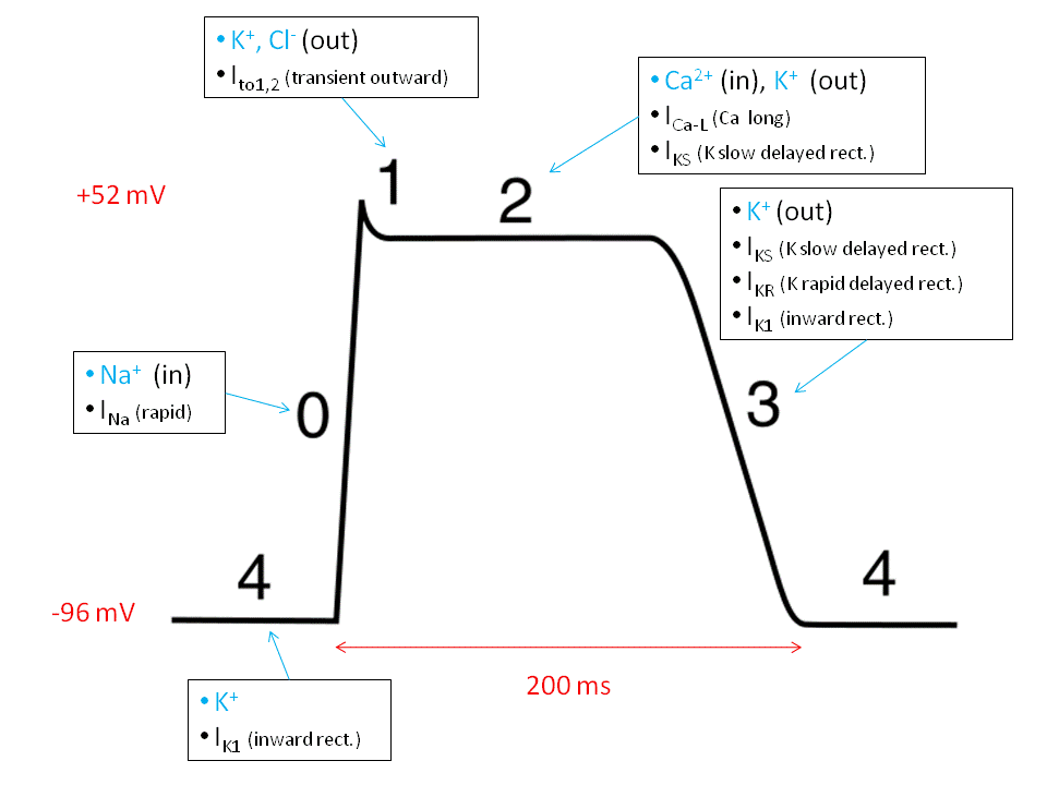
Ventricular action potential, by "Sylvia3" via Wikimedia Commons

Ventricular action potential, by “Sylvia3” via Wikimedia Commons

The ventricular contracting action potential is shown in the above figure. Since resting ventricular potential is around -90 mV, the fast Na+ channel opens to initiate a fast depolarization. Then the fast channel closes but the slow Na+–Ca++ channels open, maintaining the potential at a plateau until they close and the K+ channels open, repolarizing the cell. This long plateau, or refractory period, of about 200 ms allows time for blood to flow and avoids premature contractions which would disrupt the synchronism of the contractions.

The cardiac cycle

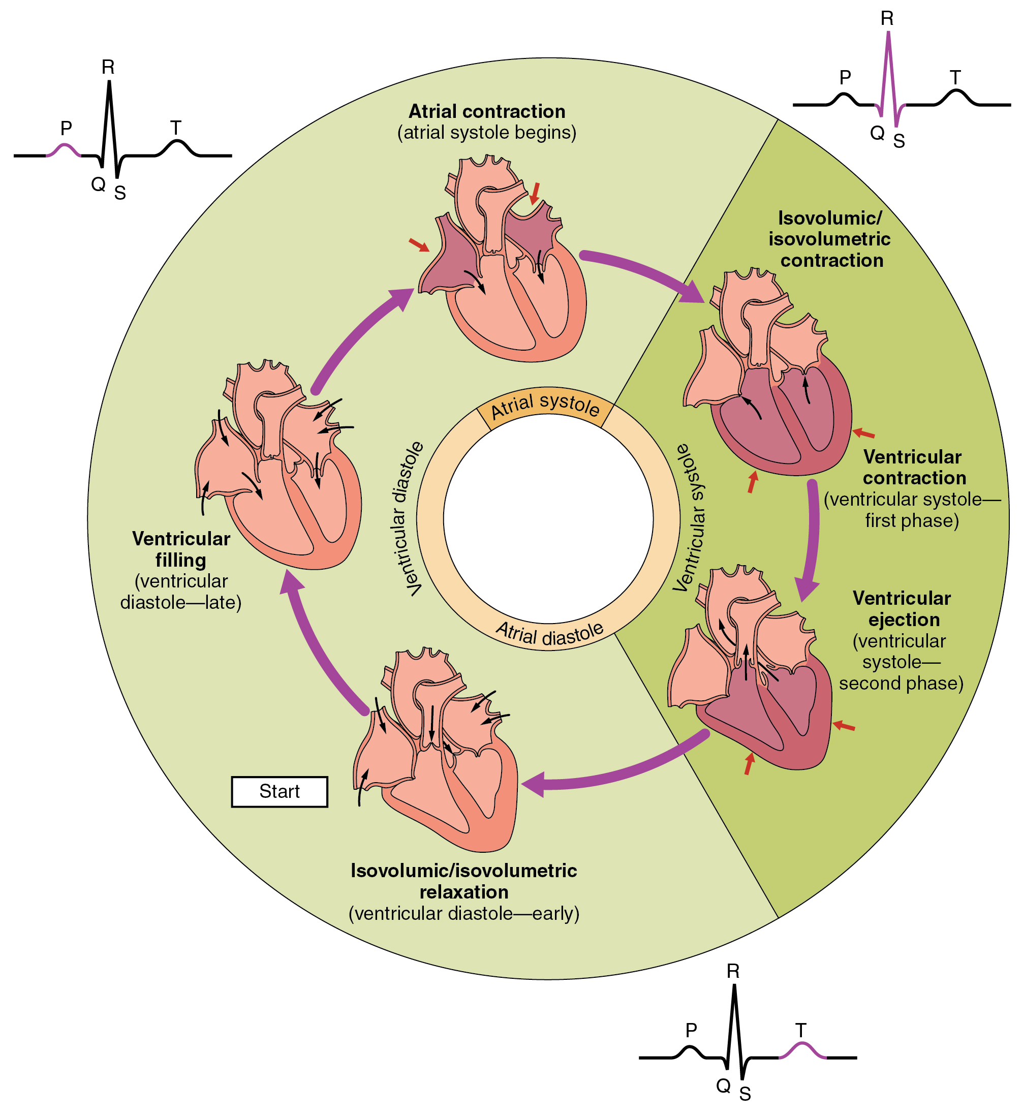
The heart’s electrical signals can be detected on the surface of the skin by an electro-cardiogram (ECG or sometimes EKG) and show the patterns of atrial and ventricular systole (contraction to pump blood out of the heart) and diastole (relaxation while the chambers refill with blood). The output shows cycles of the heart’s functioning over several steps, called the cardiac cycle. It goes like this:

  1. All chambers are relaxed (diastole) and blood flows into them, filling them to about 70-80% of their capacity. Valves keep the blood from flowing in the wrong direction.
  2. Depolarization in the SA node starts a series of action potentials which cause atrial contraction (systole) which forces the rest of the blood into the ventricles, filling them completely. This is shown as what is called the P wave on the ECG output. The signal moves down to the AV node, where there is a delay. After about 100ms,the atria relax and enter their diastole phase. They have now completed their activity for this cycle.
  3. The action potential moves down through the bundle branches and through the Purkinje fibers, launching ventrical systole. The signal reaches the right ventricle first, so it starts to contract slightly before the left. The increased pressure closes the tricuspid and mitral valves to the atria and opens the pulmonary and aortic exit valves (semilunar valves). Blood flows out to the lungs and the body. This stage shows up on the ECG as the QRS complex and lasts about 270ms. Blocks in the branches which impede the signal for whatever reason alter the form of the QRS signal.
  4. The ventricles relax into diastole at the T wave. Ventrical diastole lasts about 430ms.
Phases of the cardiac cycle, from Openstax College

Phases of the cardiac cycle, from Openstax College

The cycle starts over – constantly, all the time we are alive, from our heart’s formation during embryonic development to our last moment of life.

The “lub-dub” sound of a heartbeat, familiar to doctors with stethoscopes and to horror-movie fans, corresponds to the opening and closing of the semilunar (aortic and pulmonary) valves and so to the beginning and the end of the ventricular systole.

Regulation of heart rate

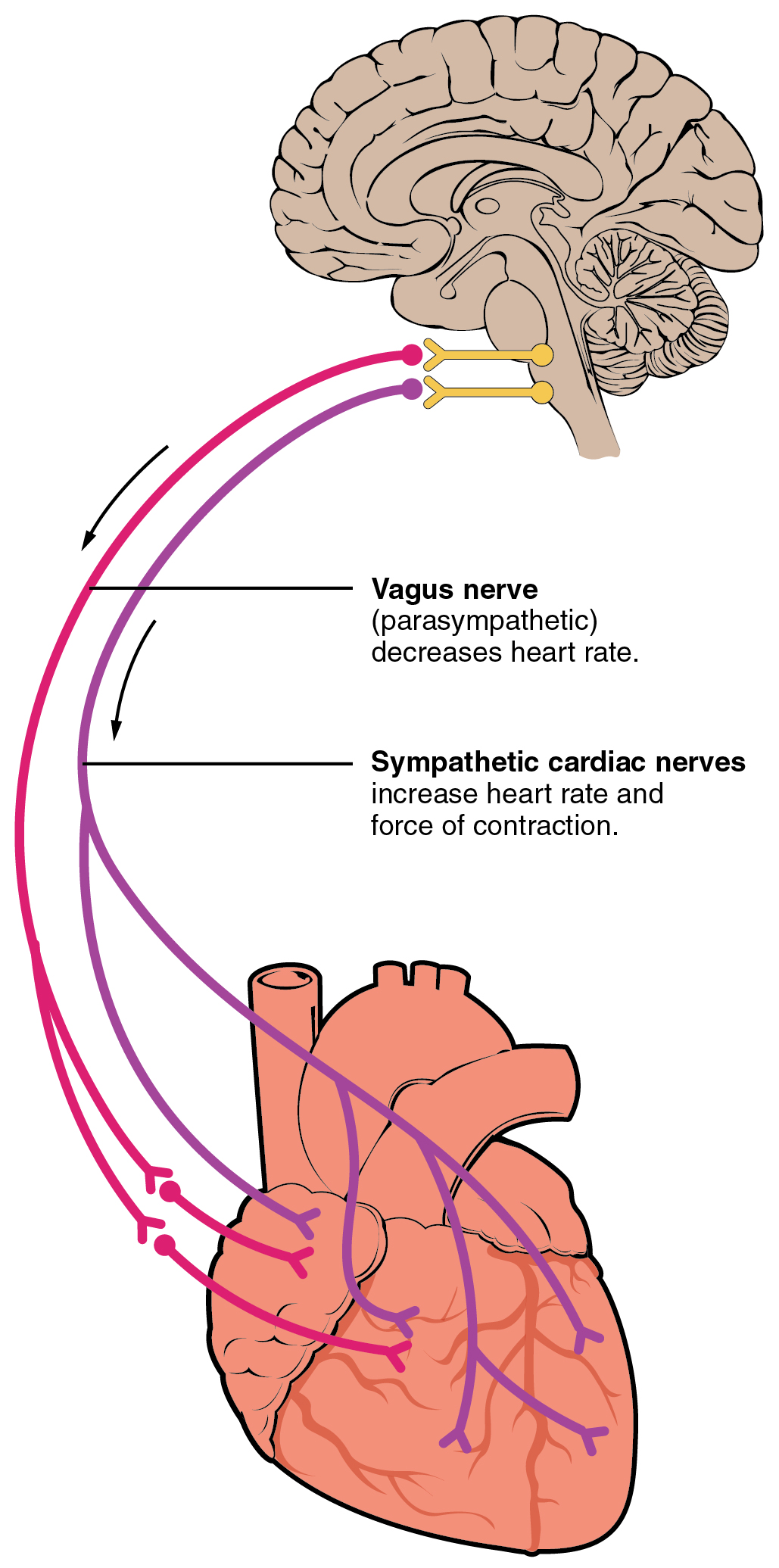
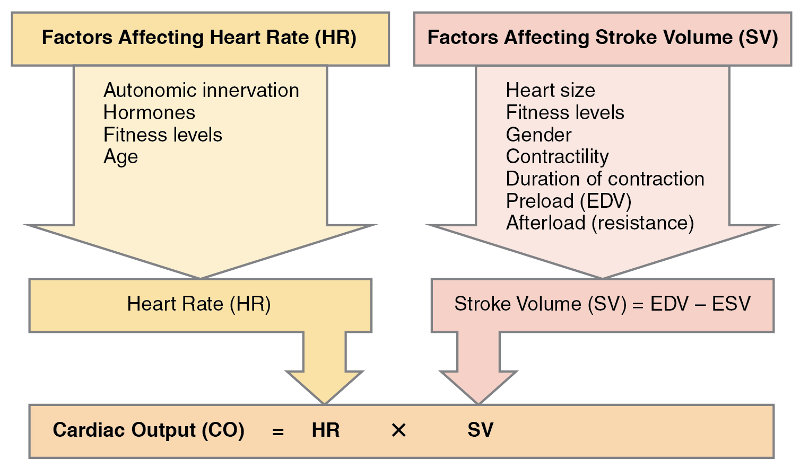
Adding up the durations of the phases indicated above gives 100+270+430 = 800ms per heartbeat, or 75bpm (beats per minute), the average rate for an adult. Although the heart itself is responsible for its beating, both the heart contraction rate (HR) and the volume of the chambers (stroke volume, SV) are influenced in several ways.

Autonomic innervation of the heart, from Openstax College

Autonomic innervation of the heart, from Openstax College

Nervous system influence on the heart originates in cardiac centers of the medulla oblongata of the hindbrain, just in front of the cerebellum. Nerve fibers from this region reach several parts of the heart, including the SA and AV nodes and both atria and ventricles. The sympathetic nervous system (e.g., “fight or flight”) releases norepinephrine (NE) which speeds up the heart rate. The parasympathetic system sends acetylcholine (Ach) along the vagus nerve which slows the heart rate. At rest, both (but mostly the parasympathetic) contribute to autonomic tone, which would otherwise be about 100bpm.

Increased physical activity detected by proprioreceptors, changes in blood pressure or flow detected by barioreceptors (stretch) or metabolic activity detected by chemoreceptors send data to the cardiac centers which then regulate heart beat and flow appropriately. Hormones, ion concentrations, temperature, pH and stress (via the limbic system) all have an influence.

Major factors influencing cardiac output, from Openstax College

Major factors influencing cardiac output, from Openstax College

Bear in mind that the heart is a muscle and, like the other muscles, needs regular exercise.

The blood

The production of blood and lymphatic system cells from hematopoietic stem cells is shown in the section on the immune system. However, the blood contains other components, listed in the table in the following figure.

Biologists distinguish four types of tissue:

  1. epithelial tissue (epithelium) covers surfaces, inside the body and out;
  2. connective tissue provides binding, support, protection and integration;2To my mind, connective tissue is “all the rest”.
  3. muscle tissue contracts and consists of three types: skeletal (voluntary), smooth and cardiac;
  4. nervous tissue propagates electrochemical signals.

Blood is considered to be connective tissue.3Obviously, it is not one of the others. Connective tissue consists of cells which are relatively far apart, compared to epithelial cells, and which are dispersed within a matrix, usually a liquid. The cells of blood, called the formed elements, float in an extracellular matrix called plasma, which is mostly water. The components of blood are shown in the following table.

Major blood components, from Openstax College

Major blood components, from Openstax College

Although blood plasma is about 92% water, it does contain three types of plasma proteins as well as small amounts of other substances.

Aside from water, the major components of the plasma are albumin and and globulins. Albumin binds to lipids and transports them through the hydrophilic plasma. It also serves to regulate osmotic pressure. Globulins, or gamma globulins, are immunoglobins, i.e., antibodies, about which more shortly.

Blood serves many purposes, but the most important (although it could not go on without the others) is the transport of oxygen, nutrients and waste matter, such as carbon dioxide.

The transport of oxygen is carried out by erythrocytes, or red blood cells (RBC), which make up 99% of the formed elements. They are so numerous, they make up approximately 25% by number of all the cells in the body.

As an erythrocyte develops in the marrow of large bones, it ejects most of its organelles, including the nucleus and the mitochondria. Without mitochondria, it is only capable of anaerobic respiration, which prevents its using up the very oxygen it is transporting. Erythrocytes are shaped like tiny disks, thinner in the middle, which enables them to fold up to fit through small openings in capillary walls. They are extraordinarily well adapted to their function. For instance, the absence of many internal organelles leaves more space for the molecule which transports oxygen: hemoglobin.

A molecule of hemoglobin is made up of four folded protein chains called globin, each one of these attached to a red pigment molecule called heme, which contains one ion of iron, Fe2+. To each iron ion, one molecule of oxygen can bind for transport, making four oxygen molecules per hemoglobin molecule. The result is called oxyhemoglobin and constitutes the bright red blood which flows in non-pulmonary arteries and pulmonary veins. The oxygen-transporting capacities of the body’s hemoglobin are quite phenomenal. One erythrocyte contains about 300 million hemoglobin molecules and so binds up to 1.2 billion oxygen molecules.

Hemoglobin molecule, modified after Openstax College

Hemoglobin molecule, modified after Openstax College

Even so, there are not too many. If the number goes down, we suffer from anemia, if not something worse. The composition of hemoglobin underlines the necessity of small amounts of trace elements like iron in the body.

The amount of oxyhemoglobin is in equilibrium with the milieu:

  • A higher concentration of oxygen leads to a greater amount of oxyhemoglobin formed;
  • a lower concentration of oxygen causes oxyhemoglobin to dissociate into oxygen and hemoglobin.

This explains why oxyhemoglobin forms in the oxygen-rich lungs and dissociates in the capillaries where oxygen is less concentrated. In addition, the presence of CO2 in the blood somewhat increases the dissociation of oxyhemoglobin.

Next, the lymphatic system.

Notes

Notes
1 Remember, veins flow towards the heart, whether they contain oxygenated blood or not.
2 To my mind, connective tissue is “all the rest”.
3 Obviously, it is not one of the others.

The endocrine system — hormones

The endocrine system, consisting of all hormone-synthesizing tissue, works together with the nervous system to regulate bodily processes. Hormones, which are endogenous (produced within the body), are far more numerous than those only produced by glands. Also, one neurotransmitter, norepinephrine, functions sometimes as a hormone and sometimes as a neurotransmitter.

Hormones are of four types:

  • lipid (or steroid) hormones – like estrogen, testosterone or cortisol, all made from a lipid, cholesterol;
  • peptide hormones – made of short chains of amino acids; include anti-diuretic hormone (ADH) and oxytocin;
  • glycoprotein (or protein) hormones – made of longer chains of amino acids; includes thyroid-stimulating hormone (TSH);
  • amine hormones – formed from the amino acids tryptophan or tyrosine; examples are melatonin, thyroid hormones, epinephrine and norepinophrine, and dopamine.

In order to have an effect, hormones must activate specific receptors on cells. A cell’s receptivity depends on the number of receptors on the cell and this can vary with time (up– or down-regulation) in response to too few or too many hormones in the blood stream, an example of the feedback control of hormonal effects.

Reception can happen in two ways. Lipid hormones are hydrophobic and therefore capable of crossing cell membranes, whereas the other three types can not. But since blood is water-based, lipid hormones must bind with a transport protein in order to travel through it. This procedure significantly increases the lifetime of the lipid-transport complex. Lipid hormones then cross the cell membrane into the cytoplasm where they bind with a receptor in the cytoplasm. The receptor-hormone complex moves into the nucleus and activates the expression of an appropriate gene to manufacture a protein which may lead to the desired result.

Binding of lipid-soluble hormones, from Openstax College

Binding of lipid-soluble hormones, from Openstax College

The other three types, non-steroid hormones (called first messengers, in this case), can travel freely through the blood, but cannot cross the cell membrane and so must bind with a receptor on the outside surface of the membrane. This begins a cascade of signals. A G protein is activated which in turn excites the so-called second messenger (usually cAMP1). Some steps later, a protein is activated by phosphorylation (addition of a phosphoryl radical) and this may lead to a cascade of many different effects, including synthesis of other products.

Binding of water-soluble hormones, from Openstax College

Binding of water-soluble hormones, from Openstax College

So steroid (lipid) hormones hitch a ride through the blood and then enter the cell to activate gene expression. Non-steroid hormones move freely through the blood, but pass the message by knocking at the cell’s door and transmitting the message to a second messenger which is already inside. 

Now let’s break it down some and take a look at some of the more important or interesting components of the endocrine system.

The following table gives some information about the principal hormones.1Table after Openstax College, http://cnx.org/contents/FPtK1zmh@8.24:4lDC0JfF@3/An-Overview-of-the-Endocrine-S, with some modifications. We will then consider some examples.

Source Hormones Class Effect

Pituitary (anterior)

Growth hormone (GH)

Protein

Growth of tissues

Pituitary (anterior)

Prolactin (PRL)

Peptide

Milk production

Pituitary (anterior)

Thyroid-stimulating hormone (TSH)

Glycoprotein

Thyroid hormone release

Pituitary (anterior)

Adrenocorticotropic hormone (ACTH)

Peptide

Adrenal cortex hormone release

Pituitary (anterior)

Follicle-stimulating hormone (FSH)

Glycoprotein

Gamete production

Pituitary (anterior)

Luteinizing hormone (LH)

Glycoprotein

Androgen production by gonads

Pituitary (posterior)

Antidiuretic hormone (ADH)

Peptide

Water reabsorption by kidneys

Pituitary (posterior)

Oxytocin

Peptide

Uterine contraction, lactation

Thyroid

Thyroxine (T4), triiodothyronine (T3)

Amine

Basal metabolic rate

Thyroid

Calcitonin

Peptide

Reduce blood Ca++ levels

Parathyroid

Parathyroid hormone (PTH)

Peptide

Increase blood Ca++ levels

Adrenal (cortex)

Aldosterone Steroid

Increase blood Na+ levels

Adrenal (cortex)

Cortisol, corticosterone, cortisone

Steroid

Increase blood glucose levels

Adrenal (medulla)

Epinephrine, norepinephrine

Amine

Fight-or-flight response

Pineal

Melatonin

Amine

Regulate circadian rhythm

Pancreas

Insulin

Protein

Reduce blood glucose levels

Pancreas

Glucagon

Protein

Increase blood glucose levels

Testes

Testoserone

Steroid

Development of male secondary sex characteristics, sperm

Ovaries

Estrogens and progesterone

Steroid

Development of female secondary sex characteristics, preparation for childbirth

Endocrine glands and cells, from Openstax College

Endocrine glands and cells, from Openstax College

The hypothalamus and the pituitary gland

The hypothalamus-pituitary complex is indeed complex. The hypothalamus, in the brain, receives signals from many parts of the nervous system. Any imbalance or non-optimal signal provokes the hypothalamus to reply through the pituitary gland, which is just below it in the brain. The hypothalamus is therefore often seen as the interface between the nervous and endocrine systems.

The pituitary gland is in two parts with rather different modes of functioning.

The posterior pituitary is really an outgrowth of the hypothalamus and is composed of nerve tissue. It stores two peptide hormones produced by the hypothalamus until the latter sends a nerve signal telling it to secrete some. Its two hormones are oxytocin, important in child bearing and rearing, and antidiuretic, or ADH (vasopresson), which signals the kidneys to control the concentration of water in the blood.2It seems like it would be easier to refer to the posterior pituitary as the lower hypothalamus instead of a gland. That it is not may be for historical reasons.

1810_Major_Pituitary_Hormones

Major pituitary hormones and their hypothalamic release hormones, from Openstax College

The anterior pituitary is composed of glandular tissue and synthesizes six main peptide hormones as well as some minor ones. It too is under the command of the hypothalamus, which communicates with it by a specific set of capillaries, the hypophyseal portal system. The hypothalamus secretes four releasing hormones and two inhibiting hormones which control secretion by the anterior pituitary. The anterior pituitary, in turn, secretes six hormones which stimulate other glands to release hormones to influence bodily functions. For instance, thyroid-stimulating hormone (TSH) stimulates the thyroid gland to produce T4 and T3 hormones and these in turn control the rates of chemical reactions elsewhere. So control goes through several steps from the hypothalamus to the anterior pituitary and on to the thyroid which then tells some other organ to do its thing.

One wonders why the two parts of the pituitary are considered to be one gland instead of two organs. Maybe this is for historical reasons.

Be that as it may, the effect of most of the pituitary hormones is to stimulate other organs to produce hormones; it is a two-step control process – three, counting the hypothalamus.

The thyroid and parathyroid glands

As shown in the above figure, the thyroid gland is located in the front of the neck. Its follicles contain thyroglobulin, which contains tyrosine amino acids. Reception of TSH from the anterior pituitary gland causes active receptors on the follicle cells to transport I (iodide) ions into the cell, where they are oxidized to I2. After peroxydase enzymes link them to tyrosine, the products finally form two hormones, triiodothyronine (T3), containing three iodines, and thyroxine (T4), containing four.3The explanation of this process given here is greatly simplified.

In a later step, low levels of T3 and T4 in the blood are detected by the hypothalamus, which releases TRH into the anterior pituitary gland, triggering it to release more TSH which in turn signals the thyroid gland to release its hormones into the blood stream. The same system allows the hypothalamus to stop TRH release, thus leading the thyroid to cease emission of its hormones. This feedback mechanism is shown in the figure.

Thyroid hormones are extremely important, having an effect on almost every physiological process in the body. They influence the body’s rate of basal metabolism, the rate of energy use when at rest. They accomplish this by modifying the amount of respiration-related enzymes within mitochondria, and by stimulating glycogen breakdown into glucose, the raw material for cellular respiration. Changing the metabolic rate also changes body temperature. Thyroid hormones also regulate rates of protein synthesis and so are important for growth, especially in young children. They eve influence mental processes.

Thyroid hormones depend on an adequate level of iodine in the body. Too much (hyperthyroidism) or too little (hypothyroidism) can lead to symptoms such as reduced mental activity or mental retardation, goiters (swollen thyroid gland), fertility or development problems

The thyroid also secretes calcitonin, a hormone whose role is to bring about a reduction in Ca++ levels in the blood. Its “other half” is PTH. PTH is secreted by two to six tiny parathyroid glands on the posterior surface of the thyroid and stimulates an increase Ca++ levels. As we have seen, this ion plays an essential role in the contraction of muscles.

Regulation of thyroid hormone levels, from Openstax College

Regulation of thyroid hormone levels, from Openstax College

The adrenal glands

The adrenal glands sit on top of the kidneys (“ad” = near, “renal” = concerning the kidney). They are in two very different parts, the adrenal cortex and the adrenal medulla. The adrenal glands are important in the body’s response to physical or psychological stress.

The adrenal cortex is the outer part of the gland and is composed of glandular tissue. It is stimulated by the hypothalamus via the anterior pituitary, called the HPA axis. The adrenal cortex plays a role in long-term stress response. Three different parts of the cortex secrete different hormones.

  • The outer cortex, the zona glomulerosa, produces mineralocorticoids, which control levels of electrolytes and liquids. The most important, aldosterone, increases the amount of Na+ ions in the blood and the volume and pressure of the blood.
  • The intermediate cortex, the zona fasciculata, produces gluticorticoids, which influence glucose metabolism. The principal one, cortisol, plays a role in stress response by making body fuel more readily available through the conversion of glycogen to glucose, the breakdown of fatty acids and glycerol, and catabolism of muscles into amino acids. It also down-regulates the immune system, which explains its use against joint inflammation, such as in hydrocortisone creams.
  • The most inner cortex, the zona reticularis, produces androgens, such as testoserone.

The adrenal medulla, in the inner part, is composed of neuroendocrine tissue and can be considered an extension of the autonomic nervous system. It is stimulated by the hypothalamus via neurons in the thoracic spinal cord, the sympathomedullary (SAM) pathway. Its major role is in short-term stress, which causes the sympathetic nervous system to alert the adrenal medulla to produce catecholamines, the hormones epinephrine (perhaps better known as adrenaline) and norepinephrine (noradrenaline). These prepare the “fight-or-flight” response by converting glycogen into glucose, raising the level of blood sugar; raising the heart rate, pulse and blood pressure; diverting blood away from digestion and other less immediate functions in order to increasing the availability of oxygen; and partially down-regulating the immune system. Thyroid hormones can up-regulate catecholamine receptors in blood vessels.

Stress response may be divided into three phases called the general adaptation syndrome, or GAS:

  1. The first stage is the alarm reaction, the short-term “fight or flight” response initiated by the adrenal cortex’s release of epinephrine or norepinephrine via the SAM pathway.
  2. If the stress continues, the stage of resistance, or adaptation, is entered. The body tries to adapt, for instance, by reducing physical activity.
  3. If the stress lasts still longer, the stage of exhaustion arrives as the adrenal cortex releases hormones via the HPA axis, as described above. Results may be depression, immune system suppression or extreme fatigue.

The pineal gland

The function of the pineal gland, a tiny gland situated inferior and slightly posterior to the thalamus, is not entirely understood. Light impinging on the retina of the eyes travels up the optic nerve to the suprachiasmatic nucleus (SCN) of the hypothalamus, which passes a signal through the spinal cord and on to the pineal gland where it inhibits the production of melatonin. This promotes wakefulness. The pineal gland therefore may influence the body’s circadian rhythms. Melatonin is used by some air travelers in an attempt to diminish the effects of changes of time-zone.

The pancreas

The pancreas is an organ with a double function. It contains exocrine cells, which release digestive enzymes directly into the small intestine. Such enzymes are essential for digestion, as we have seen in the chapter on digestion.

The pancreas also contains small structures call pancreatic islets which secrete hormones into the blood, The islets contain four types of cells.

  • alpha cells produce glucagon;
  • beta cells produce insulin;
  • delta cells secrete somatostatin, which prevents simultaneous production of glucagon and insulin;
  • PP cells produce pancreatic polypeptide hormone which plays a role in appetite and feelings of satiety.

We have already seen the important role played by glucagon and insulin in respectively raising and lowering glucose levels in the blood. They accomplish this by modulating glucose absorption and subsequent energy production by the cells (cellular respiration); the storage of glycogen in the liver (glycogenolysis); and the conversion of amino acids and glycerol into glucose (glyconeogenesis).

Endocrine functions of other organs

Other organs with endocrine functions are indicated in the table. In addition, there are a number of organs with other primary functions, but which also secrete hormones, including the heart and the skeleton. The following table covers these organs.

Organ

Major hormones

Effects

Heart

Atrial natriuretic peptide (ANP)

Reduce blood volume, pressure and Na+ concentration

Gastrointestinal tract

Gastrin, secretin, cholecystokinin (CCK)

Aid digestion, buffer stomach acids

Gastrointestinal tract

Glucose-dependent insulinotropic peptide (GIP), glucagon-like peptide 1 (GLP-1)

Stimulate pancreatic beta cells to release insulin

Kidneys

Renin

Stimulate aldosterone release

Kidneys

Calcitriol

Aid absorption of Ca++

Kidneys

Erythropoietin

Trigger red blood-cell formation in bone marrow

Skeleton

Fibroblast growth factor 23 (FSF23)

Inhibit calcitriol production, increase phosphate excretion

Skeleton

Osteocalcin

Increase insulin production

Adipose tissue

Leptin

Promote satiety signals

Adipose tissue

Adiponectin

Reduce insulin resistance

Skin

Cholecalciferol

Modified to form vitamin D

Thymus (+ other organs)

Thymosins

Aid in T-lymphocyte production, more…

Liver

Insulin-like growth factor-1

Stimulate bodily growth

Liver

Angiotensinogen

Raise blood pressure

Liver

Thrombopoetin

Cause increase in platelets

Liver

Hepcidin

Blocks iron release into bodily fluids

Another communication pathway is the circulatory system.

Notes

Notes
1 Table after Openstax College, http://cnx.org/contents/FPtK1zmh@8.24:4lDC0JfF@3/An-Overview-of-the-Endocrine-S, with some modifications.
2 It seems like it would be easier to refer to the posterior pituitary as the lower hypothalamus instead of a gland. That it is not may be for historical reasons.
3 The explanation of this process given here is greatly simplified.

More physiology — communication, circulation, immunity

The body as communications and control network

Chemical and electrochemical communications between regions and organs of the body take place constantly.

We have seen feedback processes which control certain reactions,

  • allosteric binding of reaction products;
  • control of lactose-digestion enzymes;
  • blood sugar levels controlled by insulin and glucagon;

as well as direct communication of muscle-control signals by the ANS.

We can anticipate the next chapter, on neuroscience, and see communications at work in other ways:

  • conveying sensory data input to the brain;
  • control of body functions by the central and peripheral nervous systems.

In addition to these two communication methods (chemical feedback and nervous-system control), another important system exists – hormones, the subject of which constitutes what has long been called the endocrine system (ES), although it is now known that hormones are also produced elsewhere.

The nervous system (NS) receives information from sensory organs dispersed throughout the body and in turn transmits information outwards, such as to a particular muscle cell. Communication takes place at speeds on the order of 100 meters/second and so can reach any part of the body in less than 0.01 seconds. The effect of the output, such as a muscular contraction, lasts only a short time, so the action is specific, fast and short-term.

Compared to this, communication by the endocrine system is slow and less specific, but durable. Endocrine communication takes place through the transmission of chemical messengers called hormones. Hormones are chemicals which are synthesized by tissues which may exist in specific organs called glands or in tissues or organs which also have other functions, like the pancreas.

Hormones travel through the blood until they encounter cells with special receptors which match the hormone. Any such cell may be affected. What happens then depends on the hormone and on the receiving cell. Hormones influence many bodily functions at different levels.

One may imagine a metaphor based on modern telecommunications and good ol’ radio. If the nervous system is a high-speed point-to-point link, then the hormonal system is a low-speed broadcast from a point to any stations which may be tuned to that frequency.

Another metaphor is more anthropomorphic. Chemicals which permit communication are like messengers and that is what they are often called. We will meet a number of them.

The ES and the NS work together to optimize functioning of body processes.

We will look at the following subjects:

The endocrine system — hormones

The human circulatory system

The immune system

The lymphatic system

Then we will move on to neuroscience — soon.


Muscles — motility and energy

The most obvious distinction between plants and animals is animals’ motility, our ability to move around whenever we feel like it.

Scientists think that the basic mechanism of motility is similar in all complex cells. This mechanism and its evolutionary origins were referred to briefly in the section on the cytoskeleton in the biochemistry chapter.

Motility of large organisms requires muscles. There are three types of muscles:

  • Skeletal muscles are attached to bones by tendons and contract to produce movement of the body. They are innervated voluntarily. (This is partly a question of definition. Even when they are innervated involuntarily, the innervation is said to be voluntary.
  • Cardiac muscles make the heart beat. In fact, the heart is pretty much a big muscle. The heart has its own clock, whose rate can be modified by the nervous system.
  • Smooth muscles line the digestive and respiratory tracts, surround sphincters and control the opening of the iris of the eye, among other things. Their action is involuntary, triggered by hormones, the ANS and local conditions.

Let’s look at those in more detail.

Skeletal muscles

Skeletal muscles do not just make joints move. Muscles are rarely completely relaxed, but maintain muscle tone, constantly contracting and relaxing by small amounts in order to stabilize joints and maintain balance. This is brought about by complex interactions with the nervous system.

During the development of skeletal muscles, many individual cells fuse together, usually retaining their nuclei and mitochondria. Multiple mitochondria are necessary in order to supply the great amounts of energy needed by muscles. Muscles which work more and contain more mitochondria are darker colored than those which contain fewer. Chicken legs serve to walk and are dark meat; chicken wings serve little purpose and are small and light-colored.

Three skeletal muscle structures, from Openstax College

Three skeletal muscle structures, from Openstax College

Looking from the outside in, muscle cells consist of numerous components, one nested within another, including three layers of connective tissue, or mysia (singular mysium).

  • An outer layer, the epimysium, holds the muscle together as it contracts and expands. It also connects to tendons.
  • Inside the epimysium are bundles of cells called fascicles, bounded by a second layer of connective tissue, the perimysium.
  • Inside each fascicle, the muscle cells themselves, called fibers, are bundled in another layer of connective tissue, the endomysium. The endomysium furnishes nutrition to the cells. Fibers can be quite long, up to 30 cm in the leg.
  • The fibers in turn are bundles of elements, the myofibrils, which run the length of the fiber.

Within the fibers, skeletal muscle cell components have special names, beginning with “sarco” (Greek for “flesh”). The cell membrane is called the sarcolemma; the cytoplasm, the sarcoplasm; and the smooth endoplasmic reticulum, the sarcoplasmic reticulum (abbreviated SR)

Muscle fibers, from Openstax College

Muscle fibers, from Openstax College

Skeletal muscles appear to be striated (or striped) because each myofibril is composed of a series of elements called sarcomeres, which are where the action is. The sarcomeres are placed end to end along the myofibril and contain two types of filaments: thin filaments called actin and thicker ones called myosin. The actin filaments are anchored to the ends of the sarcomeres (called Z disks or Z lines) and do not meet in the middle. The myosin filaments are positioned at the mid-point of the sarcomere, called the M line, and each end is bound to the nearest Z disc by elastic bands of the protein titin, or connectin, which act like springs, allowing the sarcomere to expand and contract while maintaining its form. The actin and myosin filaments overlap partially.

The myosin filaments are composed of multiple myosin molecules wound up together. Each molecule has a head on one end and this protrudes outward from the axis of the filament. Each head has a binding site for actin on the end and another one for ATP. In fact, the myosin head functions as an ATPase enzyme and can cleave ATP to recover the energy in it. The energy is then available for powering muscular contraction.

The actin filaments, consisting similarly of wound-up cables of actin molecules, contain binding sites for the myosin heads. When the muscle is relaxed, these sites are blocked by the protein tropomyosin, which spirals around the actin and is in turn is bound with another protein, troponin.

Thick and thin muscle filaments, from Openstax College

Thick and thin muscle filaments, from Openstax College

The neuromuscular junction (NMJ) is the point at which the motor neuron from the peripheral nervous system innervates the fiber Each fiber is connected to one branch of a motor neuron axon1An axon is the output arm of a neuron. This will be discussed in some detail in the neuroscience chapter.. If more than one fiber is connected to a neuron, the set of fibers for that neuron constitutes a motor unit. This allows for precise innervation of muscle fibers: the smaller the motor unit, the more precise the control. In the thousands of muscle fibers which move our eyeballs, a motor unit is made up of only about six fibers, making for great precision of movement. In contrast, in our back or thigh muscles, a motor unit may include thousands of muscle fibers.

When the NMJ receives an action potential, it releases the neurotransmitter acetylcholine, abbreviated Ach. Reception of Ach on the fiber receptors brings about the propagation of an action potential along the sarcolemma and through so-called T tubules to the SR, which opens calcium channels and releases Ca++ into the cell.

In more detail, the Ach receptors contain Ach-gated cation channels which open when Ach binds to them. Since the cell is normally at a negative voltage relative to its surroundings, opening the channel lets positive ions flow through the membrane and into the cell, causing it to depolarize (become less negative). This causes the opening of voltage-gated sodium channels, so Na+ enters the cell and brings about an action potential. (For more information on action potentials, see the biochemistry chapter.) When the action potential crosses the T tubules and reaches the SR, the SR releases Ca++ ions into the cell. The Ca++ binds to the troponin, which in turn causes tropomyosin to re-configure in such a way that the myosin binding sites on the actin are unblocked.

What happens next is described by the sliding-filament model of muscular contraction. (Some details of this model are still hypothetical.)

Muscle fiber contraction, from Openstax College

Muscle fiber contraction, from Openstax College

  1. The myosin heads already are bound to ADP and Pi. As the Pi is released, the myosin heads bind strongly to the actin, creating cross-bridges between the two filaments.
  2. The myosin heads then execute the power stroke: They bend toward the M-line (therefore in opposite directions on each side of the M-line), dragging the actin filaments with them (about 10 nm), increasing the overlap of the two filaments and compacting the sarcomere and, hence, the muscle.
  3. The myosin head now can bend no more. ATP binds to it and causes it to detach from the actin. The head, using energy from conversion of the ATP to ADP and Pi, returns to its neutral position. It now is “cocked” and ready to launch another power stroke, As long as the binding sites on the actin are unblocked the cycle can repeat.

Eventually, the original Ca++ is stored back into the SR by a membrane pump. Then the cycle stops until more Ca++ unblocks the actin binding sites.

Sliding-filament steps, from Openstax College

Sliding-filament steps, from Openstax College

In the sarcomere, the myosin heads drag the actin filaments along, step by step, as they overlap and the sarcomere shortens.

Sliding filaments, from Openstax College

Sliding filaments, from Openstax College

Energy for muscle action

Muscles need energy in order to function. This is why skeletal muscles have multiple mitochondria, energy factories. Energy can come from different sources and the muscles which use different sources differ in their type and function.

When a muscle is at rest, excess ATP produced may be transferred for storage to creatine phosphate (or phosphocreatine) in the muscle. This molecule stores energy in its phosphate bonds and can give it back very quickly, but can not store more than about 15 seconds worth of energy. So it is a fast, but short-duration source of energy.

Energy can also be obtained without the need of oxygen by anaerobic respiration through the conversion of glucose into ATP by the process of glycolysis, which we have already considered in some detail. This procedure is slower and, as we have seen, relatively inefficient.

After this, if oxygen still is not available, fermentation, already discussed, can convert pyruvate into lactic acid with a small gain in energy.

If oxygen is present, then aerobic respiration (Krebs cycle + oxidative phosphorylation), which is very efficient, provides energy. But it does require a regular supply of oxygen and is slower than glycolysis. So intense energy needs by muscles will first be supplied by phosphocreatine and then by glycolysis.

When muscles need ATP from aerobic respiration but the oxygen supply is short, then there is what is called an oxygen debt, which makes us breathe hard after an intense muscular effort.

Cardiac muscles

Heart muscles are striated like skeletal muscles, but are shorter and only have one nucleus per cell, although they still have many mitochondria. They are multi-branched and the branches are interconnected through intercalated discs. Gap junctions between fibers allow rapid transmission of action potentials for coordination of activity throughout the heart. The heart has its own clock and is not controlled by the nervous system.

Muscle contraction occurs through a sliding- filament scheme similar to the one in skeletal muscles. But cardiac muscles are autorhythmic: Their contractions are controlled by pacemaker cells within the heart itself, although the ANS2The autonomic nervous system, remember? may signal the heart to speed up or slow down. The pacemaker cells cause a group of cells, called a functional syncytium, to auto-excite and create an action potential. This subject will be covered in the next chapter.

Smooth muscles

Smooth muscles are not striated and contain no sarcomeres. They occur in many organs and body structures, including the digestive, urinary, respiratory, circulatory and reproductive systems, so they are quite wide-spread. They are fusiform in shape – thick in the middle and tapered at the ends. Their action is involuntary, triggered by hormones, the ANS and local conditions.

They too contract through a system similar to the sliding-filament scheme of skeletal muscles, but with important differences. Smooth muscles possess no troponin-tropomyosin complex. In a process beginning with Ca++ ions binding to a protein called calmodulin, the myosin heads are activated by being phosphorylated. The heads crawl along the actin thin filament, which is attached at its ends to dense bodies, the smooth-muscle equivalent of Z discs. But this pulls on a network of intermediate filaments, which in turn contracts the whole fiber along its length, causing it to get shorter as it swells up in the middle.

So muscles can contract because of energy from oxidative respiratioin (mostly) which uses input from food which comes from … plants — by photosynthesis.

Notes

Notes
1 An axon is the output arm of a neuron. This will be discussed in some detail in the neuroscience chapter.
2 The autonomic nervous system, remember?